
Glutathione (GSH): Redox Chemistry, Ferroptosis and Compartment Transport Research
By the TWO+DOS Research Team · Published 2026-08-13
For research use only. Not for human or veterinary use. Not for diagnostic or therapeutic use.
Glutathione is a tripeptide of glutamate, cysteine and glycine that serves as the dominant low-molecular-weight thiol inside animal cells, present at roughly 0.5 to 10 mM. Glutathione carries a free cysteine sulfhydryl group, and the reversible oxidation of that thiol to the disulfide GSSG underlies most of the chemistry studied in the laboratory.
This overview collects what peer-reviewed sources report for the molecule: verified chemical identifiers and computed properties, the two-enzyme synthesis route and its regulation, the role of the glutathione pool in ferroptosis research, the transporters that move glutathione between organelles, and results published between 2012 and 2026 in cell-free systems, cell lines, yeast and rodent models. All material described here is for laboratory research use only.
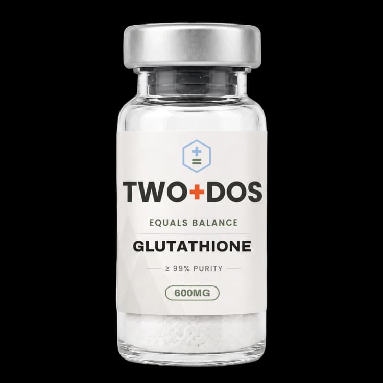
Chemical and physical properties of Glutathione
| Compound class | Endogenous thiol tripeptide |
|---|---|
| Residue order | Glutamate, cysteine, glycine, read from the amino terminus |
| Distinguishing bond | First amide forms at the glutamate γ-carboxyl, not the α-carboxyl |
| Reactive group | Free cysteine sulfhydryl, oxidized reversibly to the disulfide GSSG |
| Monoisotopic mass | 307.0838 Da (PubChem CID 124886) |
| Computed XLogP | −4.5 (PubChem), among the most hydrophilic values for a small peptide |
| Topological polar surface area | 160 Ų (PubChem computed) |
| Hydrogen bonding | 6 donors and 8 acceptors; 9 rotatable bonds (PubChem computed) |
| Reported intracellular range | Roughly 0.5-10 mM, the highest small-molecule thiol pool in animal cells |
| Reported cytosolic share | About 85-90% of the cellular pool, the balance in mitochondria, nucleus, peroxisomes and ER |
What is the chemical structure of glutathione?
Glutathione is the tripeptide γ-L-glutamyl-L-cysteinylglycine, recorded in PubChem under CID 124886 with the molecular formula C10H17N3O6S and a molecular weight of 307.33 g/mol. Glutathione differs from an ordinary tripeptide because its first amide bond forms at the γ-carboxyl of glutamate rather than at the α-carboxyl used by ribosomal peptide synthesis.
That single bond placement has a large consequence. Standard aminopeptidases and endopeptidases read α-linkages, so the γ-glutamyl bond is not a substrate for them, and the tripeptide survives in the extracellular space where an ordinary tripeptide would be cleaved in minutes. Only γ-glutamyltransferase, an ectoenzyme anchored on the outer face of the plasma membrane, hydrolyses that bond, which makes the enzyme the gatekeeper of extracellular glutathione turnover.
The computed physicochemical profile matches a highly polar zwitterionic solid. PubChem lists an XLogP of −4.5, a topological polar surface area of 160 Ų, six hydrogen-bond donors, eight acceptors and nine rotatable bonds, and a monoisotopic mass of 307.0838 Da. Two free carboxylates, one primary amine, two amide nitrogens and one thiol account for that hydrogen-bonding density and for the compound's solubility in water rather than in organic solvent.
The chemistry that matters experimentally sits on the cysteine sulfhydryl. Two molecules of the reduced form (abbreviated GSH) oxidize to one molecule of glutathione disulfide (GSSG), and the ratio between the two is the readout most laboratories use to describe intracellular redox state. Reported figures place more than 98 percent of the cellular pool in the reduced form under unstressed conditions, so measurable movement of the GSSG fraction is a sensitive signal.
How do cells build and regenerate glutathione?
Glutathione is assembled in the cytosol by two ATP-dependent enzymes acting in sequence. Glutamate-cysteine ligase joins glutamate and cysteine through the γ-carboxyl bond, and glutathione synthetase then adds glycine. Glutamate-cysteine ligase is the rate-limiting step, and cysteine availability rather than enzyme abundance frequently sets the ceiling on how much glutathione a cell can make.
Glutamate-cysteine ligase is a heterodimer of a catalytic subunit, GCLC, and a modifier subunit, GCLM, and both genes carry antioxidant response elements read by the transcription factor NRF2. Cysteine enters most cells as cystine through the system xc− antiporter, built from SLC7A11 and SLC3A2, which exports glutamate in exchange. Laboratory work blocks the pathway at two points: buthionine sulfoximine inhibits glutamate-cysteine ligase directly, while erastin blocks cystine import upstream.
Regeneration runs through glutathione reductase, a flavoenzyme that reduces GSSG back to two GSH using NADPH supplied largely by the pentose phosphate pathway. Morgan and colleagues showed in yeast that this is not the only route: cytosolic GSSG concentration stayed tightly controlled during oxidative challenge because ABC-C transporter Ycf1 moved the disulfide rapidly into the vacuole, and Trx2 and Grx2 acted as efficient backup systems to glutathione reductase. Biosensor measurements using Grx1-roGFP2 place the cytosolic glutathione redox potential in the region of −310 to −320 mV, considerably more reducing than earlier estimates based on whole-cell extracts.
Degradation proceeds by two separate routes. Outside the cell, γ-glutamyltransferase removes the glutamate and dipeptidases split the remaining cysteinylglycine, releasing amino acids for reuptake in what is classically described as the γ-glutamyl cycle. Inside the cell, the enzyme CHAC1 cleaves glutathione directly in the cytosol, and its transcriptional induction is one of the mechanisms by which cells lower their own glutathione pool during stress signalling.
Why is glutathione central to ferroptosis research?
Glutathione supplies the reducing equivalents for glutathione peroxidase 4, the selenoprotein that converts lipid hydroperoxides in membranes to the corresponding alcohols. When glutathione falls, glutathione peroxidase 4 loses its substrate, lipid hydroperoxides accumulate, and cells undergo ferroptosis, an iron-dependent death route distinct from apoptosis and necrosis.
The link was built in two steps. Dixon and colleagues reported in 2012 that erastin kills cells by inhibiting cystine uptake through system xc−, that the resulting death is iron-dependent and morphologically unlike apoptosis, necrosis or autophagy, and that ferrostatin-1 blocks it. Yang and colleagues then profiled twelve ferroptosis-inducing compounds and separated them into a class that depletes glutathione and thereby inactivates glutathione peroxidases, and a class that inhibits glutathione peroxidase 4 directly. Across 177 cancer cell lines, diffuse large B cell lymphomas and renal cell carcinomas were identified as the lineages most sensitive to that axis.
A 2025 report in Cell Chemical Biology complicated the assumption that glutathione is the only reductant the enzyme will accept. N-acetyl-l-cysteine restores the intracellular cysteine pool, but N-acetyl-d-cysteine, which cannot be converted into cysteine, blocked ferroptosis just as strongly. The authors reported that N-acetyl-l-cysteine, N-acetyl-d-cysteine and cysteine all act as reducing substrates for glutathione peroxidase 4, that only the enzyme itself was required for the effect while system xc−, glutathione biosynthesis and ferroptosis suppressor protein 1 were dispensable, and that a broad range of thiols including β-mercaptoethanol served the same function in vitro.
Work published the same year showed that the size of the total glutathione pool, and not just the GSSG to GSH ratio, carries information. Ju and colleagues reported that induction of CHAC1 shrank the pool, lowered protein S-glutathionylation and worsened ferroptosis in erastin-challenged cell lines and in hepatocytes under acetaminophen challenge, while CHAC1 deficiency enlarged the pool and attenuated hepatocyte ferroptosis.
How is glutathione distributed between cellular compartments?
Glutathione is synthesized only in the cytosol, so every other compartment depends on transport. Roughly 85 to 90 percent of the cellular pool sits in the cytosol, with the remainder in mitochondria, the nuclear matrix, peroxisomes and the endoplasmic reticulum, and each of those compartments maintains its own ratio of reduced to oxidized glutathione.
The mitochondrial carrier was identified in 2021. Wang and colleagues used organellar proteomics and metabolomics to name SLC25A39 as the transporter responsible for mitochondrial glutathione import in mammalian cells. Loss of SLC25A39 lowered mitochondrial glutathione without changing whole-cell glutathione, cells lacking both SLC25A39 and its paralogue SLC25A40 showed defects in the activity and stability of iron-sulfur cluster proteins, and mitochondrial import proved necessary for proliferation in vitro and for red blood cell development in mice.
A 2023 report in Science described how that carrier is tuned. When mitochondrial glutathione falls, the protease AFG3L2 stops degrading SLC25A39, so the carrier accumulates and import rises, and a putative iron-sulfur cluster in the matrix-facing loop of SLC25A39 is required for the response. The result couples iron status and glutathione status through a single sensing element rather than through separate pathways.
The endoplasmic reticulum runs the opposite arrangement, holding a higher GSSG to GSH ratio than the cytosol to support disulfide bond formation in secretory proteins. A 2026 study in Nature Cell Biology developed rapid immunopurification of the endoplasmic reticulum, combined the resulting proteome and metabolome with CRISPR screening, and identified SLC33A1 as the major exporter of oxidized glutathione from that compartment. A liposome assay confirmed direct GSSG transport, cryogenic electron microscopy structures and molecular dynamics simulations resolved the binding site and the residues required for transport, and loss of the exporter shifted protein disulfide isomerases toward their oxidized forms, triggering endoplasmic reticulum stress and dependence on the associated degradation pathway.
Transport also happens between cells. A 2025 Redox Biology study reported that Cx43 and Cx50 connexin hemichannels in lens epithelial cells open under mechanical stimulus and release glutathione, that Cx43 hemichannels showed higher opening efficiency and greater transport capacity than Cx50, and that fibre cells took the released glutathione back up through Cx50 hemichannels activated by fluid flow shear stress, forming an intercellular relay in a tissue with no vasculature of its own.
What is protein S-glutathionylation and how is it studied?
Glutathione forms mixed disulfides with protein cysteine residues, a reversible post-translational modification called S-glutathionylation. Glutathione attached this way alters the charge and steric environment of the modified cysteine, and glutaredoxins remove it again, so the modification behaves as a reversible switch rather than as terminal oxidative damage to the protein.
How glutaredoxins achieve selectivity was addressed in a 2024 Redox Biology study from Lang and colleagues. Class I glutaredoxins can work through a monothiol route, using a single glutathionylated active-site cysteine, or a dithiol route, forming an intramolecular disulfide between the active-site and resolving cysteines. The authors reported that S-glutathionylated glutaredoxins react rapidly with a wide range of thiols and that the second glutathione site lacks specificity for the reduced form, whereas the slower intramolecular disulfide reduction depends on specific interactions with both carboxylate groups of glutathione. The dithiol mechanism, on that reading, is what makes the enzyme selective for glutathione rather than for thiols generally.
Quantitative redox proteomics is the standard way the modification is mapped across a proteome. In the 2025 CHAC1 study, that approach identified S-glutathionylated proteins whose modification tracked pool size, and singled out ADP-ribosylation factor 6. Lower S-glutathionylation of ARF6 reduced its abundance in lysosomes, which increased transferrin receptor localization at the cell membrane and raised transferrin uptake, connecting a glutathione-dependent modification to the iron loading that ferroptosis requires.
What have 2025 and 2026 studies reported about glutathione?
Glutathione research published in 2025 and 2026 has moved beyond the intracellular antioxidant frame toward transport, compartment-specific pools and extracellular catabolism. Three findings illustrate the shift: mitochondrial import as a requirement for metastatic colonization, endoplasmic reticulum disulfide export as a determinant of protein maturation, and extracellular glutathione acting as an amino acid reservoir for tumours.
The metastasis result came from a 2025 Cancer Discovery study using mitochondrial metabolomics to compare primary and metastatic breast cancer cells. Mitochondrial glutathione accumulated during lung metastasis, driven by elevated SLC25A39 expression. Loss of the carrier impaired metastatic colonization in genetic screens, cell line models and xenograft models without affecting primary tumour growth, the requirement was specific to early colonization, and it operated independently of the carrier's canonical antioxidant role. CRISPR activation screens identified the stress-induced transcription factor ATF4 as a bypass that restored metastatic potential in carrier-deficient cells.
The 2026 Nature study reversed the usual assumption about which glutathione pool matters. Depleting intracellular glutathione did not alter tumour growth, while extracellular glutathione proved highly abundant in the tumour microenvironment. Supplying glutathione rescued cancer cell survival and growth under cystine-deficient conditions, and that rescue depended on the catabolic activity of γ-glutamyltransferases. Pharmacological blockade of γ-glutamyltransferase activity stopped the breakdown of circulating glutathione, lowered tumour cysteine levels and slowed tumour growth, framing the tripeptide as a circulating store of cysteine, glutamate and glycine rather than only as an intracellular reductant.
A 2025 review in Cancer Letters places these threads in one picture. It describes glutathione as a cofactor for glutathione peroxidase 4 in the conversion of lipid peroxides to non-toxic lipid alcohols, notes that raised glutathione synthesis and glutathione peroxidase 4 activity in cancer cells lower their ferroptosis susceptibility, and lists NFE2L2/NRF2, TP53, NF-κB, Hippo and mTOR as the signalling pathways through which glutathione status feeds back into cell behaviour.
Summary of published research
Findings below are reported as published by the cited authors, in the model systems they used. They describe laboratory research, and none of them characterize use in humans.
Dixon SJ, Lemberg KM, Lamprecht MR, et al. Ferroptosis: an iron-dependent form of nonapoptotic cell death. Cell (2012)
- Model system
- cancer cell lines and primary neuronal culture
- Conditions
- erastin challenge; iron chelation; ferrostatin-1 counter-challenge; glutamate-induced neuronal death
- Reported finding
- Erastin was reported to inhibit cystine uptake through the cystine/glutamate antiporter system xc−, compromising the glutathione-dependent antioxidant system and triggering an iron-dependent, nonapoptotic death route named ferroptosis that was morphologically and biochemically distinct from apoptosis, necrosis and autophagy, and that ferrostatin-1 blocked in both cancer cells and glutamate-challenged neurons.
Morgan B, Ezeriņa D, Amoako TN, Riemer J, Seedorf M, Dick TP. Multiple glutathione disulfide removal pathways mediate cytosolic redox homeostasis. Nature Chemical Biology (2013)
- Model system
- yeast (Saccharomyces cerevisiae) with compartment-targeted fluorescent redox probes
- Conditions
- genetically encoded roGFP2-based probes; oxidative challenge; deletion of glutathione reductase and candidate backup pathways
- Reported finding
- Compartment-specific probe measurements placed the cytosolic glutathione redox potential roughly 100 mV more reducing than previously reported, cytosolic glutathione disulfide concentration remained tightly controlled during oxidative challenge because the ABC-C transporter Ycf1 moved it rapidly into the vacuole, and Trx2 and Grx2 were identified as efficient backup systems to glutathione reductase.
Yang WS, SriRamaratnam R, Welsch ME, et al. Regulation of ferroptotic cancer cell death by GPX4. Cell (2014)
- Model system
- cancer cell line panel (177 lines) and xenograft models
- Conditions
- twelve ferroptosis-inducing compounds versus eleven compounds acting by other mechanisms; targeted metabolomic profiling and chemoproteomics
- Reported finding
- One compound class depleted glutathione and thereby inactivated glutathione peroxidases, while a second class inhibited glutathione peroxidase 4 directly; manipulating the enzyme altered sensitivity to all twelve ferroptosis inducers and none of the eleven controls, and profiling across 177 cancer cell lines identified diffuse large B cell lymphomas and renal cell carcinomas as the most susceptible lineages.
Wang Y, Yen FS, Zhu XG, et al. SLC25A39 is necessary for mitochondrial glutathione import in mammalian cells. Nature (2021)
- Model system
- mammalian cell lines and mouse
- Conditions
- organellar proteomics and metabolomics; single and combined loss of SLC25A39 and SLC25A40; proliferation and erythroid development readouts
- Reported finding
- SLC25A39 was identified as the carrier regulating glutathione transport into mitochondria; its loss lowered mitochondrial glutathione abundance without changing whole-cell glutathione, combined loss with the paralogue SLC25A40 produced defects in the activity and stability of iron-sulfur cluster proteins, mitochondrial import was required for proliferation in vitro and for red blood cell development in mice, and glutathione availability negatively regulated SLC25A39 protein abundance.
Liu Y, Liu S, Tomar A, et al. Autoregulatory control of mitochondrial glutathione homeostasis. Science (2023)
- Model system
- mammalian cell lines, isolated mitochondria
- Conditions
- glutathione depletion; AFG3L2 protease manipulation; mutation of the matrix-facing loop of SLC25A39
- Reported finding
- A feedback loop was described in which glutathione depletion releases SLC25A39 from degradation by the protease AFG3L2, allowing compensatory increases in mitochondrial glutathione uptake, and a putative iron-sulfur cluster in the matrix-facing loop of SLC25A39 was reported to be essential for the response, linking iron status and glutathione import through one sensing element.
Lang L, Reinert P, Diaz C, Deponte M. The dithiol mechanism of class I glutaredoxins promotes specificity for glutathione as a reducing agent. Redox Biology (2024)
- Model system
- cell-free (purified recombinant glutaredoxins)
- Conditions
- kinetic comparison of reduction of S-glutathionylated glutaredoxins versus intramolecular glutaredoxin disulfides across a panel of thiol reductants
- Reported finding
- S-glutathionylated glutaredoxins reacted rapidly with a wide range of thiols and the second glutathione binding site showed no specificity for the reduced tripeptide, whereas the slower reduction of the intramolecular disulfide depended on specific interactions with both carboxylate groups of glutathione, indicating that the dithiol mechanism is what confers selectivity for glutathione as the reductant.
Ju Y, Zhang Y, Tian X, et al. Protein S-glutathionylation confers cellular resistance to ferroptosis induced by glutathione depletion. Redox Biology (2025)
- Model system
- multiple cell lines and mouse hepatocytes in vivo
- Conditions
- erastin-induced ferroptosis; acetaminophen challenge in hepatocytes; CHAC1 deficiency and overexpression; quantitative redox proteomics
- Reported finding
- Upregulation of the glutathione-degrading enzyme CHAC1 shrank glutathione pools, lowered protein S-glutathionylation and worsened ferroptosis, while CHAC1 deficiency enlarged the pools, raised S-glutathionylation and attenuated hepatocyte ferroptosis; reduced S-glutathionylation of ADP-ribosylation factor 6 lowered its lysosomal abundance, increasing transferrin receptor surface localization and transferrin uptake.
Zheng J, Zhang W, Ito J, et al. N-acetyl-l-cysteine averts ferroptosis by fostering glutathione peroxidase 4. Cell Chemical Biology (2025)
- Model system
- cell lines and cell-free enzyme assays
- Conditions
- N-acetyl-l-cysteine versus N-acetyl-d-cysteine; genetic loss of system xc−, glutathione biosynthesis, glutathione peroxidase 4 and ferroptosis suppressor protein 1; in vitro reducing-substrate panel
- Reported finding
- N-acetyl-d-cysteine, which cannot be converted into cysteine, blocked ferroptosis as strongly as the l-isomer, and all three of N-acetyl-l-cysteine, N-acetyl-d-cysteine and cysteine acted as reducing substrates for glutathione peroxidase 4; only the enzyme itself was required for the effect, and a broad range of reducing substrates including β-mercaptoethanol supported its activity in vitro.
Wang G, Quan Y, Ma B, et al. Mechano-activated connexin hemichannels mediate intercellular glutathione transport and support lens redox homeostasis. Redox Biology (2025)
- Model system
- lens epithelial and fibre cells
- Conditions
- mechanical stimulation and fluid flow shear stress; comparison of Cx43 and Cx50 hemichannel activity; oxidative challenge
- Reported finding
- Cx43 and Cx50 hemichannels in lens epithelial cells released glutathione in response to mechanical stimuli, Cx43 hemichannels showed higher opening efficiency and greater transport capacity than Cx50, and glutathione released by epithelial cells was taken up by fibre cells through Cx50 hemichannels activated by fluid flow shear stress, forming an intercellular relay that lowered oxidative stress.
Yeh HW, DelGaudio NL, Uygur B, et al. Mitochondrial Glutathione Import Enables Breast Cancer Metastasis via Integrated Stress Response Signaling. Cancer Discovery (2025)
- Model system
- breast cancer cell lines, mouse metastasis models and xenograft models
- Conditions
- mitochondrial metabolomics comparing primary and metastatic cells; SLC25A39 loss; CRISPR activation screening; hypoxic culture
- Reported finding
- Mitochondrial glutathione accumulated during lung metastasis through elevated SLC25A39 expression, loss of the carrier impaired metastatic colonization without affecting primary tumour growth, the requirement was specific to early colonization and independent of the canonical antioxidant role, and CRISPR activation screens identified ATF4 as a bypass that restored metastatic potential in carrier-deficient cells.
Hecht F, Zocchi M, Tuttle ET, et al. Catabolism of extracellular glutathione supplies cysteine to support tumours. Nature (2026)
- Model system
- cancer cell lines and mouse tumour models
- Conditions
- depletion of intracellular glutathione; cystine-deficient culture with glutathione supplementation; pharmacological blockade of γ-glutamyltransferase activity
- Reported finding
- Depletion of intracellular glutathione did not alter tumour growth while extracellular glutathione was highly abundant in the tumour microenvironment; supplying glutathione rescued cancer cell survival and growth under cystine-deficient conditions in a manner dependent on γ-glutamyltransferase catabolic activity, and blocking that activity stopped breakdown of circulating glutathione, lowered tumour cysteine levels and slowed tumour growth.
Liu S, Gad M, Li C, et al. SLC33A1 exports oxidized glutathione to maintain endoplasmic reticulum redox homeostasis. Nature Cell Biology (2026)
- Model system
- mammalian cell lines, cell-free liposome transport assay and structural analysis
- Conditions
- rapid immunopurification of the endoplasmic reticulum with proteome and metabolome profiling; CRISPR screening; proteoliposome transport assay; cryogenic electron microscopy and molecular dynamics
- Reported finding
- SLC33A1 was identified as the major exporter of oxidized glutathione from the endoplasmic reticulum, loss of the transporter caused GSSG accumulation in that compartment, a liposome assay demonstrated direct GSSG transport, structures and simulations resolved the binding site and transport-critical residues, and an imbalanced GSSG to GSH ratio shifted protein disulfide isomerases toward oxidized forms while inducing endoplasmic reticulum stress and dependence on the associated degradation pathway.
What laboratory handling information is published?
Glutathione is supplied as a lyophilized powder, and its computed profile explains that format: an XLogP of −4.5, a topological polar surface area of 160 Ų and six hydrogen-bond donors describe a strongly hydrophilic solid that dissolves in aqueous buffer rather than in organic solvent. Two free carboxyl groups make aqueous solutions acidic, and published methods commonly buffer them before use in cell culture.
The free thiol is the reactive liability. Sulfhydryl groups oxidize to disulfide in air, and the rate rises with pH and with trace transition-metal contamination, so working solutions are normally prepared fresh, kept cold and handled under conditions that limit the interval between preparation and measurement. Published protocols also note that an aqueous solution left standing will accumulate the disulfide GSSG, which is the same species used analytically to report oxidation, so mishandling and biology are indistinguishable in the resulting number.
Analytical practice is well documented. A validated reversed-phase HPLC method published in Molecules quantifies the reduced form as its o-phthaldialdehyde adduct with fluorescence detection at 350 nm excitation and 450 nm emission, reporting a limit of detection of 0.34 µM and a limit of quantification of 1.14 µM for GSH, and 0.26 µM and 0.88 µM respectively for GSSG, with linearity across 0.1-4 mM for the reduced form. The same method blocks free thiol with 40 mM N-ethylmaleimide before measuring the disulfide, holds samples at 4 °C throughout, and limits total preparation time to 10-12 minutes to keep autoxidation from inflating the GSSG figure.
Identity confirmation follows standard practice for a small peptide: electrospray mass spectrometry against a monoisotopic mass of 307.0838 Da, giving a singly protonated ion near m/z 308.09, with purity reported by reversed-phase HPLC on the lot certificate of analysis. Lyophilized material is normally kept frozen, sealed against moisture and protected from light.
This material is offered for laboratory research use only. It is not a drug, not a food, and not intended for human or veterinary use.
Frequently asked research questions
What is the difference between GSH and GSSG?
GSH is the reduced tripeptide carrying a free cysteine sulfhydryl group. GSSG is glutathione disulfide, formed when two GSH molecules link through that sulfur. Reported figures place more than 98 percent of the cellular pool in the reduced form under unstressed conditions, and the ratio between the two is the measurement most laboratories use to describe intracellular redox state.
Why is the gamma linkage in glutathione significant?
The first amide bond forms at the glutamate side-chain carboxyl rather than at the α-carboxyl used in ribosomal protein synthesis. Standard peptidases do not recognize that geometry, so the tripeptide persists extracellularly where an ordinary tripeptide would be cleaved. Only γ-glutamyltransferase hydrolyses the bond, which is why that ectoenzyme controls extracellular glutathione turnover.
How is glutathione connected to ferroptosis?
Glutathione is the reducing substrate for glutathione peroxidase 4, which converts membrane lipid hydroperoxides to lipid alcohols. Compounds that block cystine import through system xc−, such as erastin, or that inhibit glutamate-cysteine ligase, such as buthionine sulfoximine, deplete the pool and leave the enzyme without substrate, so lipid hydroperoxides accumulate and iron-dependent ferroptosis follows.
Which transporters move glutathione between compartments?
Four appear repeatedly in the recent literature. SLC25A39 imports glutathione into mitochondria, with SLC25A40 as a paralogue and the protease AFG3L2 tuning its abundance. SLC33A1 was identified in 2026 as the major exporter of oxidized glutathione from the endoplasmic reticulum. Connexin hemichannels Cx43 and Cx50 move it between lens cells, and the ABC-C transporter Ycf1 moves the disulfide into the yeast vacuole.
What model systems dominate the glutathione literature?
Four recur across the citations above: cell-free enzyme and liposome assays for transport and reduction kinetics, yeast with genetically encoded roGFP2-based probes for compartment redox potential, cancer cell line panels for ferroptosis sensitivity, and mouse models covering erythroid development, hepatocyte challenge and metastatic colonization.
Glutathione at TWO+DOS
TWO+DOS supplies Glutathione as a research-use-only compound, third-party tested, with certificates of analysis emailed immediately on request.
View the Glutathionelisting →Related research overviews
References
- PubChem CID 124886: Glutathione compound summary
- Regulation of ferroptotic cancer cell death by GPX4 (Cell 2014)
- Ferroptosis: an iron-dependent form of nonapoptotic cell death (Cell 2012)
- SLC25A39 is necessary for mitochondrial glutathione import in mammalian cells (Nature 2021)
- Autoregulatory control of mitochondrial glutathione homeostasis (Science 2023)
- The dithiol mechanism of class I glutaredoxins promotes specificity for glutathione as a reducing agent (Redox Biol 2024)
- Protein S-glutathionylation confers cellular resistance to ferroptosis induced by glutathione depletion (Redox Biol 2025)
- N-acetyl-l-cysteine averts ferroptosis by fostering glutathione peroxidase 4 (Cell Chem Biol 2025)
- Mitochondrial Glutathione Import Enables Breast Cancer Metastasis via Integrated Stress Response Signaling (Cancer Discov 2025)
- Catabolism of extracellular glutathione supplies cysteine to support tumours (Nature 2026)
- SLC33A1 exports oxidized glutathione to maintain endoplasmic reticulum redox homeostasis (Nat Cell Biol 2026)
- Measurement of Glutathione as a Tool for Oxidative Stress Studies by High Performance Liquid Chromatography (Molecules 2020)
For research use only. Not for human or veterinary use. Not for diagnostic or therapeutic use.