
Oxytocin: Nonapeptide Chemistry, OXTR Structure and Magnesium-Dependent Activation
By the TWO+DOS Research Team · Published 2026-08-13
For research use only. Not for human or veterinary use. Not for diagnostic or therapeutic use.
Oxytocin is a cyclic nonapeptide hormone of the neurohypophysial family, assembled as Cys-Tyr-Ile-Gln-Asn-Cys-Pro-Leu-Gly-NH2 and closed by a disulfide bridge between cysteines 1 and 6. PubChem lists CID 439302 with the molecular formula C43H66N12O12S2 and an average molecular weight of 1007.2 g/mol. The sequence differs from arginine vasopressin at only two positions.
That two-residue separation from a closely related hormone, combined with an unusual requirement for a divalent metal ion at the receptor, defines most of what makes the molecule interesting to structural and analytical chemists. This overview collects what peer-reviewed sources report on the peptide, its receptor, the allosteric partners that govern binding, the kinetics measured in circulation, and the analytical methods used to quantify it.
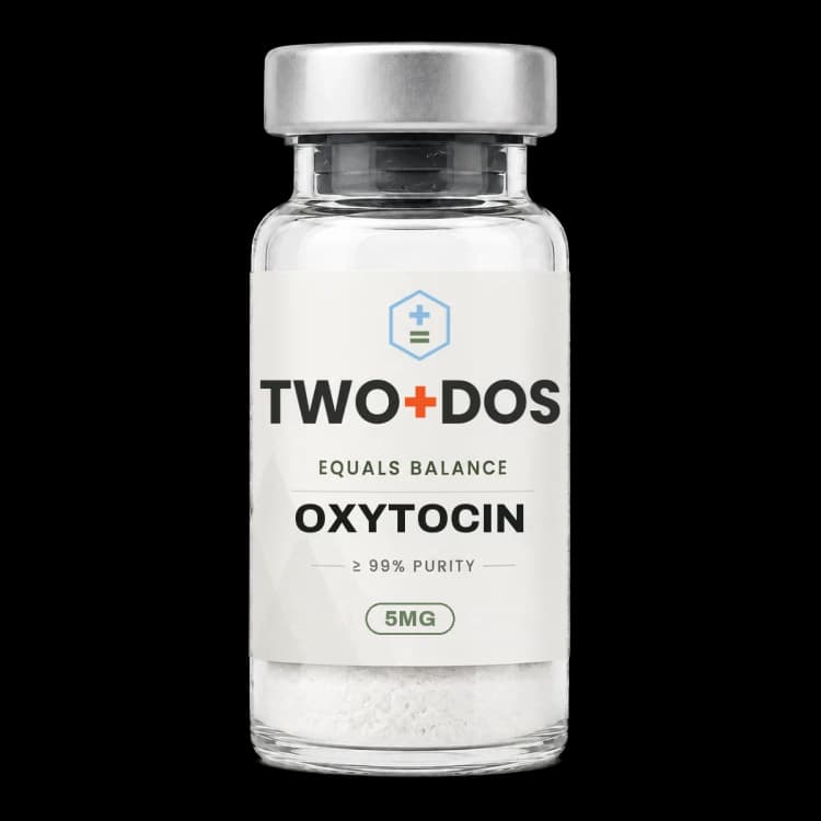
Chemical and physical properties of Oxytocin
| Compound class | Cyclic nonapeptide of the neurohypophysial (oxytocin/vasopressin) hormone family |
|---|---|
| Ring architecture | 20-membered macrocycle closed by a Cys1-Cys6 disulfide, with an acyclic Pro-Leu-Gly-NH2 tripeptide tail |
| Average molecular weight | 1007.2 g/mol for C43H66N12O12S2 (PubChem CID 439302) |
| Monoisotopic mass | 1006.43646 Da (PubChem computed) |
| Computed XLogP | -2.6 (PubChem), consistent with a strongly hydrophilic lyophilized solid |
| Topological polar surface area | 450 Ų computed, with 12 hydrogen-bond donors and 15 acceptors |
| Divergence from vasopressin | Two substitutions only: isoleucine at position 3 and leucine at position 8, where arginine vasopressin carries phenylalanine and arginine |
| Cognate receptor | OXTR, a 389-residue class A G protein-coupled receptor (UniProt P30559) coupling principally to Gq/11 |
| Biosynthetic precursor | OXT gene product oxytocin-neurophysin 1 proprotein, 125 residues (UniProt P01178) |
| Potency convention | Historically expressed in International Units traceable to the WHO International Standard held by NIBSC as code 76/575 |
How is the chemistry of Oxytocin defined?
Oxytocin carries a 20-membered macrocycle formed by the Cys1-Cys6 disulfide, with a flexible Pro-Leu-Gly-NH2 tail projecting from the ring. PubChem computes an XLogP of -2.6, a topological polar surface area of 450 Ų, twelve hydrogen-bond donors and fifteen acceptors, a profile consistent with a highly water-soluble lyophilized solid.
The relationship to arginine vasopressin is closer than the divergent physiology suggests. Both peptides share the disulfide-bridged ring and the C-terminal glycinamide, and the two molecules differ at positions 3 and 8 alone. One of the older systematic names for the compound, 3-L-isoleucine-8-L-leucine vasopressin, encodes that difference directly. Cross-reactivity at the two receptor families follows from the shared scaffold and constrains assay design.
Biosynthesis proceeds through a single precursor. The human OXT gene encodes a 125-residue oxytocin-neurophysin 1 proprotein catalogued as UniProt P01178, from which the nonapeptide is excised and C-terminally amidated. Because the free peptide and its carrier protein arise from one transcript, immunoassays that fail to distinguish the mature amidated nonapeptide from precursor fragments have been a recurring source of disagreement in the measurement literature.
Divalent metals bind the peptide itself, not only the receptor. Park and Heffern surveyed that chemistry in the European Journal of Inorganic Chemistry in 2026, describing distinct coordination modes for Cu2+, Zn2+, Mg2+ and Ca2+ and the conformational rearrangements each imposes on the macrocycle. Their review frames metal binding as an experimental variable relevant to biosensor construction and formulation chemistry alike.
How does Oxytocin engage the oxytocin receptor?
Oxytocin signals through OXTR, a 389-residue class A G protein-coupled receptor catalogued in UniProt as P30559. Receptor occupancy couples principally to Gq/11, activating phospholipase C-β, hydrolyzing phosphatidylinositol 4,5-bisphosphate into inositol trisphosphate and diacylglycerol, and mobilizing intracellular calcium in the smooth muscle and neuronal populations where the receptor is expressed.
The first atomic-resolution view arrived in 2020. Waltenspühl and colleagues solved the human receptor by X-ray diffraction at 3.2 Å in complex with retosiban, a non-peptidic OXTR-selective antagonist, and deposited the coordinates as PDB entry 6TPK. Two features of that map proved consequential: an extrahelical cholesterol molecule wedged between helices IV and V, and a conserved coordination site for Mg2+ shared across neurohypophysial receptors.
The active state followed two years later. Meyerowitz and colleagues determined the wild-type human receptor bound to the peptide and to miniGq/i by cryo-electron microscopy at 2.9 Å, released as PDB entry 7RYC. The structure showed the agonist and the receptor jointly forming a Mg2+ coordination complex, and the bound peptide disrupting transmembrane helix 7. Functional assays in the same work traced full agonism to that TM7 disruption and partial agonism by analogs to its absence.
Binding affinities place the peptide firmly at its own receptor while leaving measurable cross-reactivity. Taylor and colleagues determined competition binding constants in Syrian hamster brain in 2020 and reported Ki 4.28 nM at OXTR against 495.2 nM at V1aR, while arginine vasopressin gave 4.70 nM at V1aR and 36.1 nM at OXTR. The selectivity pattern they measured resembled human receptors more closely than rat receptors, a species caveat that matters when rodent data are compared across labs.
Why do magnesium and cholesterol change Oxytocin receptor binding?
Oxytocin binding at OXTR depends on two allosteric partners that sit outside the peptide binding pocket: a divalent magnesium ion and membrane cholesterol. Both were identified structurally rather than inferred, and both raise agonist affinity, which makes buffer composition and membrane lipid content decisive variables in any receptor assay.
Cholesterol occupancy was mapped computationally in 2025. Marlow and colleagues combined molecular docking with molecular dynamics in the Biophysical Journal and resolved cholesterol sites on both agonist-bound and antagonist-bound receptor states, with differing residence times between them. Allosteric network analysis singled out one site on the extracellular leaflet between TM4 and TM5, and two intracellular-leaflet sites between TM2, TM3 and TM4 and between TM4 and TM5, as the routes carrying signal to the orthosteric pocket. Several predicted positions coincided with unassigned density in an experimental cryo-EM map.
The magnesium site received comparable scrutiny in 2026. Musiani and colleagues, writing in the Journal of Inorganic Biochemistry, applied molecular dynamics with a purpose-built force field for the divalent ion and predicted four water molecules completing the octahedral coordination shell that experimental maps leave incomplete. Their simulations placed the peptide C-terminal region and the receptor N-terminal region simultaneously near the ion, and modelled intracellular loop 3, unresolved in every deposited structure, in two conformations linking TM5 and TM6.
Cation dependence turns out to be a family-level switch rather than a quirk. The 2022 cryo-EM work identified a single cation-coordinating residue whose identity determines whether a given vasopressin-family receptor requires a divalent ion at all, an observation that rationalizes why some members of the family are magnesium-sensitive and others are indifferent.
What do pharmacokinetic studies report for Oxytocin?
Oxytocin disappears from the circulation on a timescale of minutes. Fabian and colleagues reported in the Journal of Physiology in 1969 a median half-life of 3.2 minutes after a bolus, rising to 4.8 minutes during continuous infusion, with metabolic clearance of roughly 1.5 litres per minute in human volunteers.
Ultrafiltration in that same 1969 work found the peptide entirely unbound in plasma, against about 30% binding for arginine vasopressin. Modern mass spectrometry has since revised the picture without softening it. Shafer and colleagues published a population model in the British Journal of Anaesthesia in 2025 built on LC-MS/MS rather than antibody capture, across two protocols in healthy adults. Systemic delivery fitted a two-compartment model with 0% bias and 18% median inaccuracy. Nasal delivery of 100 µg fitted the same model shape but with 9% median bias and 47% median inaccuracy, and nasal bioavailability came out at 0.7%.
Enzymatic clearance runs largely through a zinc metalloenzyme. Insulin-regulated aminopeptidase, the enzyme also referred to as oxytocinase, hydrolyzes the peptide while resisting the structurally related angiotensin IV. Hanževački and colleagues explained that discrimination with QM/MM molecular dynamics in 2025, attributing cleavage to a stabilizing interaction between the sigma hole of the N-terminal disulfide and the lone pair of the scissile nitrogen, and attributing resistance in the competing substrate to geometric strain that destabilizes the tetrahedral intermediate.
Direct imaging of nasal delivery reached humans in 2025. Winterdahl and colleagues synthesized [13N]oxytocin and scanned six volunteers by PET/MRI, reporting high nasal-cavity uptake within the first five minutes, time-activity curves indicating tracer presence in brain regions between 25 and 45 minutes, and uptake that was low, variable between individuals, and without a clear relationship to the quantity presented. The nasal cavity was the dosimetry-limiting organ, and the authors concluded the tracer is not presently suited to central receptor imaging by that route.
Where has Oxytocin receptor expression been mapped?
Oxytocin receptor expression has been mapped at single-transcript resolution. Ryu and colleagues applied RNAscope to whole female and male mouse brains and published the resulting atlas in eLife in 2026, identifying the nuclei carrying the highest transcript density for the peptide and for its receptor across the intact brain.
That atlas presents the receptor as a peripheral as well as a central target, noting direct actions on bone remodeling and skeletal calcium mobilization. Receptor identity and ligand identity are separate questions. Inada and colleagues reported in Nature Communications in 2025 that vasopressin neurons of the paraventricular hypothalamus signal partly through oxytocin receptors expressed in the anterior commissure and medial nuclei of the preoptic area in male mice. Those receptor-expressing neurons received input from both peptide populations, making the receptor a convergence point rather than a private line for one ligand.
Peripheral receptor pools complicate the nasal-delivery literature further. Sun and colleagues reported in the European Journal of Pharmacology in 2026 that mouse nasal epithelium harbours receptor-expressing neurons, and that volatilized peptide exposure raised Fos expression in the epithelium, main olfactory bulb mitral cells, piriform cortex and cortical amygdala. Lavage with a receptor antagonist abolished the zone-preference behaviour in male mice, and the effect was absent in receptor knockout animals, placing part of the response at the epithelium itself.
What have 2025 and 2026 publications added to the Oxytocin record?
Oxytocin publications from 2025 and 2026 cluster into four groups: computational dissection of the magnesium and cholesterol allosteric sites, bivalent ligand chemistry aimed at receptor homodimers, analytical methods pushing quantification into the picogram range, and human protocols probing receptor blockade and nasal biodistribution rather than agonist delivery.
The homodimer chemistry is the most pharmacologically specific of these. Busnelli and colleagues reported in Biochemical Pharmacology in 2026 that homobivalent compounds built from two modified analogs, joined by C8 and C10 spacers, engage both protomers of a receptor homodimer through an interface involving transmembrane domains 1 and 2 and helix 8. BRET biosensors showed the two optimal-spacer compounds activating Gi2 and Gi3 in addition to Gq, while sparing Gi1, GoA and GoB, and recruiting β-arrestin 1 alongside β-arrestin 2. The same pair had previously shown roughly 100-fold and 40-fold potency gains over the native peptide in mouse and zebrafish social-behaviour assays.
Human work has moved toward blocking the receptor rather than supplying the ligand. Fitzek and colleagues ran a randomized, double-blind, placebo-controlled crossover provocation study published in the Journal of Headache and Pain in 2026, giving the receptor antagonist atosiban as a 6.75 mg bolus followed by 54 mg over three hours to sixty participants across three groups. Migraine-like attacks in the primary group occurred in 6 of 20 with antagonist against 4 of 20 with placebo (p = 0.75), and the healthy comparison group showed increased temporal artery diameter and middle cerebral artery flow velocity where the migraine groups did not.
Analytical sensitivity has kept pace. Zhang and colleagues published an LC-MS/MS urine method in Biomedical Chromatography in 2026 using mixed-mode solid-phase extraction and serially coupled columns, reaching a lower limit of quantification of 1 pg/mL with extraction recovery above 80%, precision under 9% and accuracy between 101% and 109% in human urine. Parallel radiochemistry appeared in Bioconjugate Chemistry in 2025, where gallium-68-labelled peptides targeting the receptor were assembled by linchpin chemistry that performs cyclization and chelator incorporation in tandem.
Summary of published research
Findings below are reported as published by the cited authors, in the model systems they used. They describe laboratory research, and none of them characterize use in humans.
Fabian M, Forsling ML, Jones JJ, Pryor JS. The clearance and antidiuretic potency of neurohypophysial hormones in man, and their plasma binding and stability. The Journal of Physiology (1969)
- Model system
- human volunteers
- Conditions
- bioassay measurement of plasma hormone levels following a single bolus and during continuous infusion; ultrafiltration for plasma protein binding; stability comparison in human plasma at 4 °C and 37 °C
- Reported finding
- Median half-life was 3.2 minutes after a bolus and 4.8 minutes during infusion, against 5.5 to 5.7 minutes for the vasopressins; metabolic clearance was 1.5 litres per minute compared with 1.0 for vasopressin, apparent volumes of distribution were about two thirds of extracellular volume, and ultrafiltration found the nonapeptide completely unbound where about 30% of arginine vasopressin was plasma-bound.
Hawe A, Poole R, Romeijn S, Kasper P, van der Heijden R, Jiskoot W. Towards heat-stable oxytocin formulations: analysis of degradation kinetics and identification of degradation products. Pharmaceutical Research (2009)
- Model system
- cell-free accelerated stability study
- Conditions
- formulations at pH 2.0, 4.5, 7.0 and 9.0 held at 40, 55, 70 and 80 °C; rate constants from RP-HPLC; characterization by HP-SEC, UV absorption and fluorescence spectroscopy; degradation products identified by ESI-MS/MS
- Reported finding
- Loss followed first-order kinetics with strong pH and temperature dependence, fastest at pH 9.0, and the Arrhenius activation barrier peaked at 116.3 kJ/mol for the pH 4.5 formulation; deamidated species, intramolecular oligosulfides and covalent aggregates were identified unequivocally, and the whole pH-dependent profile was described by the Arrhenius equation.
Taylor JH, McCann KE, Ross AP, Albers HE. Binding affinities of oxytocin, vasopressin and Manning compound at oxytocin and V1a receptors in male Syrian hamster brains. Journal of Neuroendocrinology (2020)
- Model system
- rodent (male Syrian hamster brain tissue)
- Conditions
- saturation binding to establish Kd for the radioligands [125I]ornithine vasotocin analogue and [125I]linear vasopressin antagonist, followed by competition binding to derive Ki at OXTR and V1aR
- Reported finding
- Each peptide bound its canonical receptor most tightly, with Ki 4.28 nM (95% CI 2.9 to 6.3) at OXTR for the nonapeptide and 4.70 nM at V1aR for arginine vasopressin, against 495.2 nM and 36.1 nM respectively at the non-canonical receptor; Manning compound gave 6.87 nM at V1aR and 213.8 nM at OXTR, and the hamster selectivity profile resembled human receptors more than rat receptors.
Waltenspühl Y, Schöppe J, Ehrenmann J, Kummer L, Plückthun A. Crystal structure of the human oxytocin receptor. Science Advances (2020)
- Model system
- X-ray crystallography of purified human receptor
- Conditions
- human OXTR in complex with the non-peptidic selective antagonist retosiban, solved at 3.2 Å and deposited as PDB entry 6TPK, combined with mutagenesis and binding experiments
- Reported finding
- The map resolved the antagonist contacts in detail and revealed an extrahelical cholesterol molecule occupying an unexpected position between helices IV and V, supplying a structural rationale for the long-documented cholesterol dependence of the receptor; the same work identified a conserved neurohypophysial-receptor-specific Mg2+ coordination site acting as a positive allosteric modulator of agonist binding.
Meyerowitz JG, Robertson MJ, Barros-Álvarez X, Panova O, Nwokonko RM, Gao Y, Skiniotis G. The oxytocin signaling complex reveals a molecular switch for cation dependence. Nature Structural & Molecular Biology (2022)
- Model system
- cryo-electron microscopy of a purified receptor-G protein complex
- Conditions
- wild-type human OXTR bound to the native nonapeptide and to miniGq/i, resolved at 2.9 Å and deposited as PDB entry 7RYC, with parallel functional assays on the receptor and on peptide analogs
- Reported finding
- Activation required both a Mg2+ coordination complex formed jointly by the peptide and the receptor and disruption of transmembrane helix 7 by the bound agonist, with TM7 disruption separating full agonism from the partial agonism shown by analogs; the identity of a single cation-coordinating residue was shown to determine whether any given vasopressin-family receptor is cation-dependent.
Marlow B, Vogel A, Kuenze G, Pankonin M, Reinhardt F, Stadler PF, Hildebrand PW, Meiler J. Cholesterol allosteric modulation of the oxytocin receptor. Biophysical Journal (2025)
- Model system
- molecular docking and molecular dynamics simulation of the human receptor in membrane
- Conditions
- cholesterol site prediction on agonist-bound and antagonist-bound receptor states, with residence-time analysis, allosteric network analysis, and comparison against density in an experimental cryo-EM map
- Reported finding
- Docking and dynamics converged on several cholesterol sites with state-dependent distributions and residence times, one of which coincided with unassigned experimental cryo-EM density; network analysis identified an extracellular-leaflet site between TM4 and TM5 and two intracellular-leaflet sites, between TM2, TM3 and TM4 and between TM4 and TM5, as the principal conduits carrying allosteric signal into the orthosteric pocket.
Hanževački M, Twidale RM, Lang EJM, Gerrard W, Wright DW, Stojević V, Mulholland AJ. Quantum Mechanics/Molecular Mechanics Simulations Distinguish Insulin-Regulated Aminopeptidase Substrate (Oxytocin) and Inhibitor (Angiotensin IV) and Reveal Determinants of Activity and Inhibition. Journal of Chemical Information and Modeling (2025)
- Model system
- QM/MM molecular dynamics of the zinc metalloenzyme insulin-regulated aminopeptidase
- Conditions
- enhanced-sampling QM/MM simulations of hydrolysis for the nonapeptide substrate against angiotensin IV, comparing tetrahedral-intermediate geometries and reaction barriers
- Reported finding
- Cleavage of the nonapeptide was stabilized by an interaction between the sigma hole of the N-terminal disulfide bond and the hybridizing lone pair of the scissile peptide nitrogen, while the competing peptide showed a larger deviation of the valine C-Cα-Cβ angle from ideal tetrahedral geometry that destabilized its intermediate, explaining why one is turned over and the other behaves as an inhibitor.
Shafer SL, Ririe DG, Miller S, Curry RS, Hsu DT, Sullivan GM, Eisenach JC. Plasma pharmacokinetics of intravenous and intranasal oxytocin in nonpregnant adults. British Journal of Anaesthesia (2025)
- Model system
- human volunteers (healthy adult men and nonpregnant women)
- Conditions
- two protocols, the first delivering 16.7 µg into the bloodstream over 1 or 10 minutes and the second 13.7 µg over 30 minutes with 100 µg intranasal on a separate day (n = 24); venous plasma quantified by LC-MS/MS alongside enzyme-linked immunosorbent assay, with parameters estimated in NONMEM
- Reported finding
- Systemic kinetics were well described by a two-compartment model with 0% bias and 18% median inaccuracy, while the nasal route showed 9% median bias and 47% median inaccuracy with bioavailability of 0.7%; mass-spectrometry values ran systematically higher than simultaneous immunoassay values, and the authors attributed inconsistent reports across the nasal literature partly to that sub-1% bioavailability and large between-subject spread.
Winterdahl M, Nielsen EN, Hansen SB, Dias AH, Vendelbo MH, Jakobsen S, Yeomans D. First-in-human intranasal [13N]oxytocin PET: evaluation of feasibility, biodistribution, and radiation dosimetry. EJNMMI Research (2025)
- Model system
- human volunteers (six healthy participants)
- Conditions
- intranasal [13N]-labelled tracer followed by whole-body or head PET/MRI, with time-activity curve extraction, trigeminal ganglion and brain region analysis, and radiation dosimetry modelling
- Reported finding
- Tracer accumulated heavily in the nasal cavity within five minutes before declining into systemic absorption, with time-activity curves indicating presence in brain regions between 25 and 45 minutes; uptake in trigeminal ganglia and brain was low and varied between individuals with no clear relationship to the quantity presented, one participant with rhinitis showed altered kinetics, and the nasal cavity was the dosimetry-limiting organ.
Zhang X, Fralick L, Ethun K. Quantitative Determination and Stability of Trace Levels of Oxytocin in Human and Rhesus Macaque Urine by LC-MS/MS. Biomedical Chromatography (2026)
- Model system
- analytical method development in human and rhesus macaque urine
- Conditions
- mixed-mode solid-phase extraction with serially coupled chromatography columns, validated per FDA guidance, including bench-top, frozen and freeze-thaw stability testing
- Reported finding
- The method reached a lower limit of quantification of 1 pg/mL with extraction recovery above 80%, precision under 9% and accuracy between 101% and 109% in human urine, and 93% to 109% accuracy with precision under 11% in macaque urine; the analyte proved stable at room temperature for 4 hours, at -80 °C for 30 days, in extracted form at 4 °C for 24 hours, and across up to three freeze-thaw cycles.
Busnelli M, Gori A, Chini B. Bivalent ligands targeting oxytocin receptor homodimers selectively activate distinct G protein and β-arrestin pathways. Biochemical Pharmacology (2026)
- Model system
- cell-based BRET biosensor assays on the human receptor
- Conditions
- a series of homobivalent compounds built from two modified analogs joined by spacers of varying length, profiled against G protein subtypes and both β-arrestin isoforms
- Reported finding
- The C8 and C10 spacer compounds, which bridge both protomers of a homodimer through an interface involving transmembrane domains 1 and 2 and helix 8, activated Gi2 and Gi3 in addition to Gq while leaving Gi1, GoA and GoB untouched, and were the only members of the series recruiting β-arrestin 1 as well as β-arrestin 2; the same pair had previously shown roughly 100-fold and 40-fold potency gains over the native peptide in mouse and zebrafish social-behaviour assays.
Musiani F, Al Sadi S, Giorgetti A, Alfonso-Prieto M, Carloni P. Structural and functional role of the magnesium ion in the human oxytocin receptor. Journal of Inorganic Biochemistry (2026)
- Model system
- molecular dynamics simulation of the human receptor-peptide complex
- Conditions
- simulations using a purpose-built force field for the divalent cation, with intracellular loop 3 modelled in two conformations because it is unresolved in every deposited experimental structure
- Reported finding
- Four water molecules were predicted to complete the octahedral coordination shell of the Mg2+ ion left incomplete by experimental maps; binding was enhanced when the peptide C-terminal region and the receptor N-terminal region approached the ion simultaneously, while activation additionally depended on the conformation of transmembrane helices 5 and 6 joined by the modelled intracellular loop 3.
What laboratory handling information is published?
Oxytocin is supplied as a lyophilized powder, and the published stability record explains why. Hawe and colleagues reported first-order loss across pH 2.0, 4.5, 7.0 and 9.0 at 40, 55, 70 and 80 °C, fastest at pH 9.0, with degradation describable by the Arrhenius equation. The activation barrier peaked at 116.3 kJ/mol for the pH 4.5 formulation.
The degradation chemistry is unusually well characterized for a peptide of this size. Wiśniewski and colleagues traced the route in Biopolymers in 2013 using HPLC, LC-MS and 13C-NMR on material held at 40 °C: β-elimination opens the disulfide to give a linear intermediate bearing an S-thiocysteine persulfide at position 6 and a dehydroalanine at position 1. That persulfide donates sulfur to an intact molecule, generating trisulfide and higher monomeric polysulfides, while the dehydroalanine hydrolyzes with loss of the N-terminal amino group to give a linear N-pyruvoylated octapeptide. Terminal products include dimers of that octapeptide linked through disulfide bridges and aldol-type condensations.
Quantification is the second recurring problem. Antibody-based assays and mass spectrometry do not agree: the 2025 population kinetics work found LC-MS/MS values systematically higher than simultaneous immunoassay values on the same samples, which is one reason older concentration figures resist direct comparison. The 2026 urine method establishes what current chromatography can reach, with a validated lower limit of quantification of 1 pg/mL and documented stability at room temperature for 4 hours, at -80 °C for 30 days and across three freeze-thaw cycles.
Metal content is a variable rather than an inert background. Because Cu2+, Zn2+, Mg2+ and Ca2+ each impose distinct coordination geometries on the macrocycle, and because Mg2+ acts as a positive allosteric modulator at the receptor, buffer composition changes both the conformational population of the peptide in solution and the affinity measured in a binding assay. Membrane cholesterol content exerts the same kind of influence on the receptor side. Reported handling parameters describe laboratory conditions only. This material is for research use, is not for human or animal consumption, and no preparation guidance is provided.
Frequently asked research questions
What is Oxytocin?
Oxytocin is a cyclic nonapeptide hormone, C43H66N12O12S2, average molecular weight 1007.2 g/mol, catalogued as PubChem CID 439302 and CAS 50-56-6. A disulfide bridge between cysteines 1 and 6 closes a 20-membered ring, leaving a Pro-Leu-Gly-NH2 tail. It acts at OXTR, a class A G protein-coupled receptor catalogued as UniProt P30559.
How does Oxytocin differ from vasopressin at the chemical level?
The two peptides share the disulfide-bridged ring, the nine-residue length and the C-terminal glycinamide, and differ at positions 3 and 8 only: isoleucine and leucine here, phenylalanine and arginine in arginine vasopressin. Competition binding in hamster brain gave Ki 4.28 nM at OXTR and 495.2 nM at V1aR for the former, and 4.70 nM and 36.1 nM for the latter.
Why does magnesium matter in Oxytocin receptor assays?
Structural work identified a coordination site for Mg2+ conserved across neurohypophysial receptors, and the 2022 active-state cryo-EM structure showed the peptide and the receptor forming that coordination complex jointly during activation. The ion behaves as a positive allosteric modulator of agonist binding, so magnesium concentration in an assay buffer is a determinant of measured affinity rather than an incidental detail.
What does the literature report about intranasal Oxytocin reaching the brain?
The evidence is mixed and the numbers are small. Population kinetics published in 2025 measured nasal bioavailability at 0.7% with 47% median inaccuracy between subjects. First-in-human PET imaging with a [13N]-labelled tracer the same year found heavy nasal-cavity accumulation, detectable but low and highly variable signal in brain regions between 25 and 45 minutes, and concluded the tracer was not suited to central receptor imaging by that route.
Is this Oxytocin material available for human use?
No. This is a research chemical supplied as lyophilized powder for laboratory research use only. It is not for human or animal consumption, has not been evaluated by the FDA for any use in people, and no preparation or delivery instructions are provided. Everything described on this page reports published findings in the model systems the cited studies used.
Oxytocin at TWO+DOS
TWO+DOS supplies Oxytocin as a research-use-only compound, third-party tested, with certificates of analysis emailed immediately on request.
View the Oxytocinlisting →Related research overviews
References
- PubChem Compound Summary for CID 439302, Oxytocin
- UniProtKB P30559: Oxytocin receptor (OXTR), Homo sapiens
- UniProtKB P01178: Oxytocin-neurophysin 1 proprotein (OXT), Homo sapiens
- RCSB PDB 6TPK: Crystal structure of the human oxytocin receptor
- RCSB PDB 7RYC: Oxytocin receptor (OTR) bound to oxytocin in complex with a heterotrimeric Gq protein
- Waltenspühl Y, Ehrenmann J, Vacca S, Thom C, Medalia O, Plückthun A. Structural basis for the activation and ligand recognition of the human oxytocin receptor. Nature Communications 2022;13:4153
- Wiśniewski K, Finnman J, Flipo M, Galyean R, Schteingart CD. On the mechanism of degradation of oxytocin and its analogues in aqueous solution. Biopolymers 2013;100(4):408-421
- Inada K, Hagihara M, Yaguchi K, Irie S, Inoue YU, Inoue T, Miyamichi K. Vasopressin-to-oxytocin receptor crosstalk in the preoptic area underlying parental behaviors in male mice. Nature Communications 2025;16:10844
- Ryu V, Gumerova AA, Pevnev G, et al. Single transcript level atlas of oxytocin and the oxytocin receptor in the mouse brain. eLife 2026;15:e95215
- Sun LH, Chan YH, Yu Z, et al. Nasal epithelial cells expressing oxytocin receptor are involved in volatilized oxytocin effects on biased and social motivations and stress-declined neural stem cell proliferation. European Journal of Pharmacology 2026;1014:178522
- Fitzek MP, Kleist P, Handtmann C, et al. Oxytocin receptor antagonism in migraine: a randomized, double-blind, placebo-controlled provocation study. The Journal of Headache and Pain 2026;27(1)
- Park J, Heffern MC. Metal Modulation of Oxytocin Structure, Function, and Receptor Interactions. European Journal of Inorganic Chemistry 2026
For research use only. Not for human or veterinary use. Not for diagnostic or therapeutic use.