
DSIP (Delta Sleep-Inducing Peptide): Nonapeptide Chemistry and Research Findings
By the TWO+DOS Research Team · Published 2026-08-13
For research use only. Not for human or veterinary use. Not for diagnostic or therapeutic use.
DSIP, or delta sleep-inducing peptide, is a nine-residue peptide with the sequence Trp-Ala-Gly-Gly-Asp-Ala-Ser-Gly-Glu and an average formula mass of 848.8 g/mol. Swiss investigators recovered the material from rabbit cerebral venous blood and reported its full characterization in 1977. The synthetic form is handled in laboratories as a lyophilized, water-soluble solid.
Fifty years of literature have not resolved what the molecule is. No gene encoding a DSIP precursor has been identified, no receptor has been cloned, and the peak of experimental work sits between 1977 and the mid-1990s. Publications from 2023 onward are dominated by engineered fusion constructs, synthetic-chemistry demonstrations and historical reviews rather than by fresh pharmacology, which is worth knowing before reading any single result as settled.
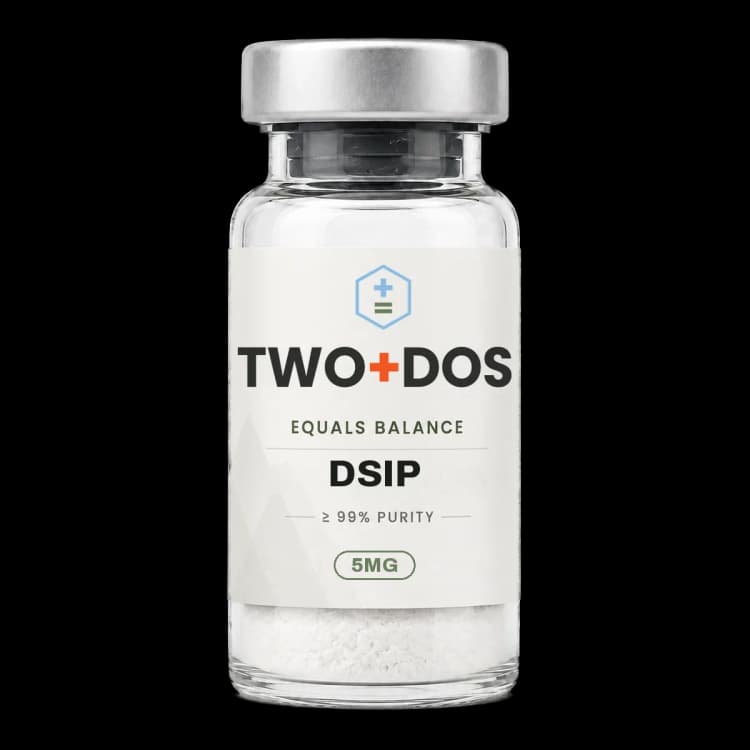
Chemical and physical properties of DSIP
| Peptide class | Linear nonapeptide. Nine residues, no cysteine, no disulfide bridge and no cyclic constraint, so identity rests entirely on sequence and mass rather than on a folded scaffold |
|---|---|
| Amino acid sequence | Trp-Ala-Gly-Gly-Asp-Ala-Ser-Gly-Glu, written WAGGDASGE in single-letter code. Glycine occupies positions 3, 4 and 8, which is why the backbone is unusually flexible for a peptide of this length |
| Molecular formula | C35H48N10O15 for the free peptide |
| Molecular mass | 848.8 g/mol average formula mass and 848.33 Da monoisotopic, so the singly protonated ion falls near m/z 849.34 in high-resolution spectra and near m/z 849.8 on an average-mass instrument |
| CAS number | 62568-57-4. The same PubChem record also carries 69431-45-4 as a secondary registry number, so both appear across supplier and reference-standard documentation |
| PubChem CID | 68816 |
| UNII | YN28Z5YZ73 |
| Nonproprietary name | Emideltide is the international nonproprietary name assigned to the nonapeptide. No medicine bearing that name has been authorized by any national regulator |
| Ionization | Three acidic groups (the aspartate side chain, the glutamate side chain and the C-terminal carboxylate) sit against a single N-terminal amine, giving a net charge near minus two at pH 7 and an isoelectric point in the acidic range. No lysine, arginine or histidine is present |
| Chromophore | Tryptophan at position 1 is the only aromatic residue and supplies essentially all absorbance at 280 nm, close to 5,500 M-1 cm-1 by standard residue additivity, which makes A280 the practical concentration check |
| Phosphorylation site | Serine 7 is the residue carried in phosphorylated form by the analogue reported as P-DSIP, and casein kinase II phosphorylates the nonapeptide in cell-free assays |
How was DSIP first identified?
DSIP was characterized in 1977 by Schoenenberger and Monnier, who reported a nonapeptide recovered from rabbit cerebral venous blood that enhanced delta and spindle activity in the electroencephalogram after intraventricular infusion. Nine synthetic peptides were compared across 58 rabbits under double-blind conditions, and only the complete nonapeptide produced the effect.
The 1977 report tested five plausible breakdown fragments spanning residues 1-8, 2-9, 2-8, 1-4 and 5-9, two nonapeptide analogues with two residues exchanged, and the tripeptide Trp-Ser-Glu. None reproduced the electroencephalographic signature. Requiring all nine residues was, at the time, the strongest argument that a discrete molecular entity rather than a degradation artefact had been isolated.
A 2025 historical review by Banks places the isolation inside a wider argument then unfolding about whether peptides could reach brain tissue at all. The same review notes that DSIP was explicitly shown not to be Pappenheimer's Factor S, the separate somnogenic material recovered from goat cerebrospinal fluid, and that a radioimmunoassay for DSIP requiring eight of its nine residues was developed to track it.
What is the published chemistry of the DSIP molecule?
DSIP carries the formula C35H48N10O15, an average mass of 848.8 g/mol and a monoisotopic mass of 848.33 Da, indexed in PubChem as CID 68816 under CAS 62568-57-4 and the nonproprietary name emideltide. Three of the nine residues are glycine, one is tryptophan, and none is cysteine.
The residue composition sets the practical analytical handles. A single tryptophan at position 1 carries the 280 nm chromophore, so ultraviolet quantitation is straightforward but sensitive to oxidation of that one residue. Two acidic side chains plus the C-terminal glutamate carboxylate against one free amine leave the molecule anionic at neutral pH, and the absence of lysine, arginine and histidine means no strong cation-exchange handle exists for purification.
Solution-state spectroscopy published in 1994 described DSIP as sitting in a dynamic equilibrium between unordered and folded states, with beta-turn conformations occupying roughly 40 percent of the population. Three glycines make that flexibility unsurprising. Phosphorylation is the one well-documented covalent modification: casein kinase II phosphorylates the peptide in cell-free assays, and serine 7 is the site carried by the analogue reported in the literature as P-DSIP.
Which mechanism and transport questions remain open?
DSIP has no identified receptor. A 2006 review in the Journal of Neurochemistry titled the peptide a still unresolved riddle precisely because neither a gene encoding a precursor nor a binding site was ever found, leaving open whether the reported activity belongs to the nonapeptide itself or to something co-purified with it in the original preparations.
Transport across the blood-brain barrier is better documented than receptor pharmacology. Passage of synthetic DSIP into rabbit brain was reported in 1977, a vascularly perfused guinea pig preparation in 1989 described uptake consistent with a high-affinity saturable carrier rather than free diffusion, and Banks records in 2025 that DSIP and its analogues crossed from blood into brain tissue and cerebrospinal fluid in intact form in rats and dogs, attributing the passage to lipid solubility.
Proposed downstream mechanisms remain descriptive. A 2026 review in the International Journal of Molecular Sciences summarizes the peptide as probably acting at multiple sites with influence over GABAergic and NMDA glutamatergic signalling. Separate rodent work on the phosphorylated analogue under simulated high altitude reported that naloxone abolished the observed changes, implicating opioid signalling. None of these accounts amounts to a defined molecular target.
What did electroencephalographic studies in animals record?
DSIP electroencephalography in rodents produced its clearest numbers with the phosphorylated analogue. A 1989 study in unrestrained rats delivered a ten-hour nocturnal intracerebroventricular infusion and recorded slow-wave activity 22 percent above baseline and paradoxical states 81 percent above baseline at 0.5 nmol, driven mainly by a greater number of episodes rather than longer ones.
The same study reported a narrow effective window. Quantities were tested from 0.025 to 25 nmol, and amounts both larger and smaller than 0.5 nmol were inactive. Potency of the phosphorylated analogue was put at five times that of the unmodified nonapeptide under matched conditions. An inverted-U response of that shape is a recurring feature of the DSIP literature and complicates comparison between studies that sampled only one level.
The most recent animal work uses engineered constructs rather than the bare peptide. A 2024 Frontiers in Pharmacology study fused DSIP to a cell-penetrating carrier sequence, expressed it in Pichia pastoris, and tested it in Kun-Ming mice made insomniac with para-chlorophenylalanine. Daily wakefulness ran at 480 plus or minus 30 minutes in controls, 720 plus or minus 45 minutes in the model group, and 500 plus or minus 32 minutes with the fusion construct.
What did controlled studies in human volunteers report?
DSIP was carried into small controlled human studies during the 1980s and early 1990s, and the results were mixed. A double-blind trial published in Neuropsychobiology in 1992 enrolled 16 adults with chronic insomnia across five laboratory nights at 25 nmol/kg, measured higher efficiency and shorter latency against placebo, and concluded that the statistically significant effects were weak.
A 1987 study in European Neurology took a different design, following fourteen middle-aged adults with chronic insomnia across seven successive nights under double-blind, placebo-controlled conditions. That report described night rest and daytime function reaching the levels of normal controls, with alertness and daytime task scores rising. The contrast between a seven-night schedule and a three-night one is the most obvious variable separating the two outcomes.
Endocrine results did not transfer from rodents to humans. A 1995 Psychoneuroendocrinology study measured corticotropin and cortisol responses during and after infusion and found them almost identical to placebo, with the authors stating explicitly that their data do not support an inhibitory role for DSIP on corticotropin and cortisol secretion in man.
What non-somnogenic effects appear in the DSIP literature?
DSIP research extends well beyond electroencephalography, and most of it sits in rodent stress models. A 1985 Neuroendocrinology study reported that corticosterone rises driven by corticotropin-releasing factor fell significantly at 5 to 30 micrograms per kilogram, while corticosterone released by direct corticotropin stimulation was unchanged, placing the effect at the pituitary rather than the adrenal cortex.
Mitochondrial work from 2003 examined isolated rat brain mitochondria by polarographic oxygen measurement. DSIP raised state 3 respiration, the respiratory control ratio and the rate of ADP phosphorylation while uncoupled respiration stayed flat, and at 120 micrograms per kilogram before a hypoxic challenge it blocked the fall in mitochondrial respiratory activity that hypoxia otherwise caused. Rat hepatocyte studies published in 2016 reported catalase and superoxide dismutase changes that varied non-monotonically across 40, 120 and 360 micrograms per kilogram.
A 2021 focal stroke study in Sprague-Dawley rats is the most rigorous recent rodent report and is notable for what it did not find. Intranasal delivery at 120 micrograms per kilogram left infarct volume at day 21 statistically indistinguishable from vehicle, 20.9 plus or minus 6.9 percent against 24.1 plus or minus 4.6 percent, while rotarod coordination in the DSIP group returned by the seventh test day. A 2026 orthopaedic review reaches the same broad conclusion for the compound class, noting that preclinical reports outnumber clinical trials by a wide margin.
Summary of published research
Findings below are reported as published by the cited authors, in the model systems they used. They describe laboratory research, and none of them characterize use in humans.
Schoenenberger GA, Monnier M. Characterization of a delta-electroencephalogram (-sleep)-inducing peptide. Proc Natl Acad Sci U S A. 1977;74(3):1282-6. (1977)
- Model system
- Rabbit, intraventricular infusion, double-blind electroencephalographic scoring
- Conditions
- Nine synthetic peptides compared across 58 rabbits including controls; neocortical and archicortical leads analysed by fast Fourier transform
- Reported finding
- Only the complete nonapeptide Trp-Ala-Gly-Gly-Asp-Ala-Ser-Gly-Glu produced significant and specific enhancement of delta and spindle patterns. Five fragment analogues spanning residues 1-8, 2-9, 2-8, 1-4 and 5-9, two substituted nonapeptides and the tripeptide Trp-Ser-Glu were all inactive.
Kimura M, Inoue S. The phosphorylated analogue of DSIP enhances slow wave sleep and paradoxical sleep in unrestrained rats. Psychopharmacology (Berl). 1989;97(1):35-9. (1989)
- Model system
- Rat, unrestrained, chronic electroencephalographic recording
- Conditions
- Ten-hour nocturnal intracerebroventricular infusion; range from 0.025 to 25 nmol examined
- Reported finding
- At 0.5 nmol the phosphorylated analogue raised slow-wave activity by 22 percent and paradoxical states by 81 percent, the increase coming mainly from a greater number of episodes. Larger and smaller quantities were inactive, and potency was reported as five times that of the unmodified nonapeptide.
Graf MV, Kastin AJ, Coy DH, Fischman AJ. Delta-sleep-inducing peptide reduces CRF-induced corticosterone release. Neuroendocrinology. 1985;41(4):353-6. (1985)
- Model system
- Rat, in vivo endocrine challenge
- Conditions
- 5 to 30 micrograms per kilogram against corticotropin-releasing factor and against direct corticotropin challenge
- Reported finding
- Corticosterone elevations driven by corticotropin-releasing factor fell significantly across the 5 to 30 microgram per kilogram range, while corticosterone released by direct corticotropin stimulation was unaffected, locating the interaction at the pituitary rather than the adrenal cortex.
Graf MV, Saegesser B, Schoenenberger GA. Degradation and aggregation of delta sleep-inducing peptide (DSIP) and two analogs in plasma and serum. Peptides. 1987;8(4):599-603. (1987)
- Model system
- Human and rat plasma and serum, cell-free biostability assay
- Conditions
- Labelled DSIP and two analogues followed by gel filtration chromatography
- Reported finding
- DSIP degraded rapidly in both human and rat blood, releasing material eluting at the retention time of free tryptophan. The two analogues broke down more slowly and instead formed complexes, which the authors advanced as one explanation for variable results between in vivo studies.
Khvatova EM, Samartzev VN, Zagoskin PP, Prudchenko IA, Mikhaleva II. Delta sleep inducing peptide (DSIP): effect on respiration activity in rat brain mitochondria and stress protective potency under experimental hypoxia. Peptides. 2003;24(2):307-11. (2003)
- Model system
- Isolated rat brain mitochondria and brain homogenate; whole-animal hypoxia
- Conditions
- Polarographic oxygen consumption; 120 micrograms per kilogram intraperitoneally before hypoxic exposure
- Reported finding
- DSIP increased the rate of phosphorylating respiration V3, the respiratory control ratio and the rate of ADP phosphorylation while uncoupled respiration remained unchanged. Pretreatment at 120 micrograms per kilogram fully blocked the hypoxia-associated reduction in mitochondrial respiratory activity.
Tukhovskaya EA, Ismailova AM, Shaykhutdinova ER, Slashcheva GA, Prudchenko IA, Mikhaleva II, Khokhlova ON, Murashev AN, Ivanov VT. Delta Sleep-Inducing Peptide Recovers Motor Function in SD Rats after Focal Stroke. Molecules. 2021;26(17):5173. (2021)
- Model system
- Sprague-Dawley rat, 90-minute intraluminal middle cerebral artery occlusion
- Conditions
- 120 micrograms per kilogram intranasally, once before occlusion and daily for seven days; ten animals with DSIP, nine with vehicle, seven sham
- Reported finding
- Infarct volume at day 21 was 20.9 plus or minus 6.9 percent with DSIP against 24.1 plus or minus 4.6 percent with vehicle, a difference that did not reach statistical significance, while rotarod coordination in the DSIP group returned by the seventh test day.
Tatsumi T, Sasamoto K, Matsumoto T, Hirano R, Oisaki K, Kanai M. Practical N-to-C peptide synthesis with minimal protecting groups. Commun Chem. 2023;6(1):231. (2023)
- Model system
- Synthetic organic chemistry, solution phase
- Conditions
- N-to-C chain elongation without elaborate condensation reagents or protecting-group manipulation
- Reported finding
- The nonapeptide DSIP was chosen as a demonstration target and assembled by direct N-to-C extension, showing that the sequence can be built without the protecting-group scaffolding that conventional C-to-N routes require.
Mu X, Qu L, Yin L, Wang L, Liu X, Liu D. Pichia pastoris secreted peptides crossing the blood-brain barrier and DSIP fusion peptide efficacy in PCPA-induced insomnia mouse models. Front Pharmacol. 2024;15:1439536. (2024)
- Model system
- Kun-Ming mouse, para-chlorophenylalanine insomnia model; 48 animals, six per group
- Conditions
- PCPA at 300 mg/kg for five consecutive days; DSIP, the carrier peptide and the DSIP-CBBBP fusion delivered intraperitoneally at 100 nM
- Reported finding
- Daily wakefulness ran at 480 plus or minus 30 minutes in controls, rose to 720 plus or minus 45 minutes in PCPA animals and fell to 500 plus or minus 32 minutes with the fusion construct, which shifted serotonin, dopamine, glutamate and melatonin measurements further than the unfused nonapeptide.
Banks WA. Synergies from a distance: Inspirations from the struggles of Dr James M Krueger. Neurobiol Sleep Circadian Rhythms. 2025;19:100114. (2025)
- Model system
- Historical review
- Conditions
- Account of radioimmunoassay development and blood-brain barrier experiments on DSIP in rats and dogs
- Reported finding
- The review records that DSIP and its analogues crossed from blood into brain tissue and cerebrospinal fluid in intact form in rats and dogs, attributes the passage to lipid solubility, and notes the peptide was shown to be distinct from Pappenheimer's Factor S.
Opp MR, Imeri L. Sleep and immune health: How dogs, goats and 'factor S' shaped a field. Neurobiol Sleep Circadian Rhythms. 2025;19:100118. (2025)
- Model system
- Narrative historical review
- Conditions
- Survey of candidate endogenous somnogenic factors from the 1970s onward
- Reported finding
- DSIP appears as one of only four candidate factors listed in the 1980 Borbely and Tobler review, alongside arginine vasotocin, Factor S and an unidentified substance, and is described as sequenced, synthesized and transported through the blood-brain barrier by the late 1970s.
Rahman OF, Lee SJ, Seeds WA. Therapeutic Peptides in Orthopaedics: Applications, Challenges, and Future Directions. J Am Acad Orthop Surg Glob Res Rev. 2026;10(1). (2026)
- Model system
- Narrative review of peptide classes in musculoskeletal research
- Conditions
- Literature survey covering wound-repair peptides, secretagogues and neuroactive compounds
- Reported finding
- The review describes DSIP as a hypothalamic neuropeptide named for promoting delta-wave activity in rodent studies, cites reports of raised endogenous growth hormone secretion and altered stress hormone responses, and concludes that the preclinical record is not matched by clinical trials.
Renke G, Chinellato L. Therapeutic Peptides in Aesthetic, Metabolic and Endocrine Conditions: Effects, Safety, Clinical Applications, and Future Perspectives. Int J Mol Sci. 2026;27(9):3890. (2026)
- Model system
- Structured review of 106 articles, weighted toward systematic reviews and randomized trials
- Conditions
- Peptides grouped by application area; DSIP covered among neuroactive compounds
- Reported finding
- The review lists DSIP as a nine-residue peptide reported in hypothalamus and cerebrospinal fluid, describes probable action at multiple sites involving GABA and NMDA glutamatergic pathways, reports breakdown by cerebrospinal-fluid peptidases with a half-life of minutes, and states the peptide was never approved and circulates only as a research compound.
What laboratory handling information is published?
DSIP is supplied as a lyophilized solid and is characterized by the same two orthogonal checks used for any small synthetic peptide: reversed-phase HPLC for purity, commonly specified at 98 percent or better in reference-standard documentation, and mass spectrometry for identity against an expected protonated ion near m/z 849.34 monoisotopic or 849.8 on an average-mass readout.
Concentration is easy to verify because the molecule carries exactly one tryptophan and no tyrosine or cysteine. Absorbance at 280 nm therefore maps to a molar absorptivity close to 5,500 M-1 cm-1 by standard residue additivity, and any drift in that figure across a stored batch points at oxidation of the single indole side chain rather than at bulk loss of peptide.
Stability in biological matrices is poor and well documented. A 1987 Peptides study following labelled material in human and rat blood found rapid breakdown with release of a species eluting at the retention time of free tryptophan, and a 2026 review reports peptidase-driven clearance from cerebrospinal fluid on a timescale of minutes. Chemical degradation in storage runs on a slower clock: the aspartyl-alanine motif at positions 5 and 6 is a classic isoaspartate-forming site, and isoaspartyl DSIP is used in the enzymology literature as a defined substrate for protein L-isoaspartyl methyltransferase, with a reported Michaelis constant of 33.36 micromolar for the yeast enzyme.
Storage practice for material of this class follows the chemistry rather than any published protocol specific to the nonapeptide: sealed and desiccated at minus 20 degrees Celsius or colder for the dry solid, protected from light because of the tryptophan chromophore, and worked up in aqueous buffer near neutral pH where the net negative charge keeps the peptide soluble. Material described here is offered for laboratory research use only. It is not a medicine, not a food, and not intended for diagnostic use or for use in humans or animals.
Frequently asked research questions
Is DSIP an endogenous peptide?
Reviews describe DSIP-like material in hypothalamus, brain tissue and cerebrospinal fluid, and a radioimmunoassay for it was developed in the late 1970s. No gene encoding a DSIP precursor has ever been identified, however, so whether the nonapeptide is a genuine gene product or a fragment of something larger remains unsettled.
Does DSIP have a known receptor?
No receptor for DSIP has been cloned or pharmacologically defined. A 2006 review in the Journal of Neurochemistry made the absence of both a gene and a binding site its central point. Recent reviews describe involvement of GABAergic and NMDA glutamatergic signalling in general terms rather than naming a target.
What is the molecular mass of DSIP?
The free peptide has the formula C35H48N10O15, an average formula mass of 848.8 g/mol and a monoisotopic mass of 848.33 Da. Mass spectrometry on the intact nonapeptide therefore looks for a singly protonated ion near m/z 849.34 at high resolution.
How does P-DSIP differ from DSIP?
P-DSIP is the analogue phosphorylated on serine at position 7, a modification that casein kinase II performs on the nonapeptide in cell-free assays. In a 1989 rat study the phosphorylated form was reported as roughly five times more potent than the unmodified peptide under matched infusion conditions.
Why is the DSIP literature considered inconsistent?
Three factors recur across the reviews: rapid breakdown in blood and cerebrospinal fluid, an inverted-U response in which quantities above and below a narrow window are inactive, and small study sizes in the human work of the 1980s and 1990s. Rodent endocrine findings also failed to reproduce in human volunteers.
DSIP at TWO+DOS
TWO+DOS supplies DSIP as a research-use-only compound, third-party tested, with certificates of analysis emailed immediately on request.
View the DSIP (5mg)listing →Related research overviews
References
- PubChem CID 68816 — delta sleep-inducing peptide (emideltide) compound record
- Schoenenberger GA, Monnier M. Characterization of a delta-electroencephalogram (-sleep)-inducing peptide. PNAS 1977
- Schoenenberger GA et al. The delta EEG (sleep)-inducing peptide (DSIP). XI. Amino-acid analysis, sequence, synthesis and activity of the nonapeptide. Pflugers Arch 1978
- Graf MV, Kastin AJ. Delta-sleep-inducing peptide (DSIP): a review. Neurosci Biobehav Rev 1984
- Kovalzon VM, Strekalova TV. Delta sleep-inducing peptide (DSIP): a still unresolved riddle. J Neurochem 2006
- Zlokovic BV et al. Saturable mechanism for delta sleep-inducing peptide (DSIP) at the blood-brain barrier of the vascularly perfused guinea pig brain. Peptides 1989
- Gray RA et al. Delta-sleep-inducing peptide: solution conformational studies of a membrane-permeable peptide. Biochemistry 1994
- Nakamura A, Shiomi H. Phosphorylation of delta sleep-inducing peptide (DSIP) by casein kinase II in vitro. Peptides 1991
- Bes F, Hofman W, Schuur J, Van Boxtel C. Effects of delta sleep-inducing peptide on sleep of chronic insomniac patients. A double-blind study. Neuropsychobiology 1992
- Schneider-Helmert D. Effects of delta-sleep-inducing peptide on 24-hour sleep-wake behaviour in severe chronic insomnia. Eur Neurol 1987
- Spath-Schwalbe E et al. Delta-sleep-inducing peptide does not affect CRH and meal-induced ACTH and cortisol secretion. Psychoneuroendocrinology 1995
- Banerjee S et al. Enzymatic attributes of an l-isoaspartyl methyltransferase from Candida utilis and its role in cell survival. Biochem Biophys Rep 2015
For research use only. Not for human or veterinary use. Not for diagnostic or therapeutic use.