
Methylcobalamin: Corrin Structure, Methionine Synthase Chemistry and Cell Trafficking
By the TWO+DOS Research Team · Published 2026-08-13
For research use only. Not for human or veterinary use. Not for diagnostic or therapeutic use.
Methylcobalamin is the methylated form of vitamin B12, a cobalt-centred corrinoid of formula C63H91CoN13O14P and molecular mass 1344.4 g/mol, catalogued under CAS 13422-55-4 and PubChem CID 10898559. A methyl group bonded directly to cobalt makes the molecule one of two organometallic cofactors found in mammalian cells.
That single bond is what separates methylcobalamin from the other cobalamins sold as research chemicals. Cyanocobalamin and hydroxocobalamin carry non-alkyl upper ligands and are photostable; methylcobalamin and adenosylcobalamin carry alkyl groups and are not. The sections below cover the architecture of the corrin ring, the catalytic cycle of methionine synthase, the intracellular route by which cells build the molecule from imported cobalamin, the distinctive chemistry of the cobalt-carbon bond, the microbial methyltransferases that use the same chemistry, and what cell-based work published since 2025 has reported.
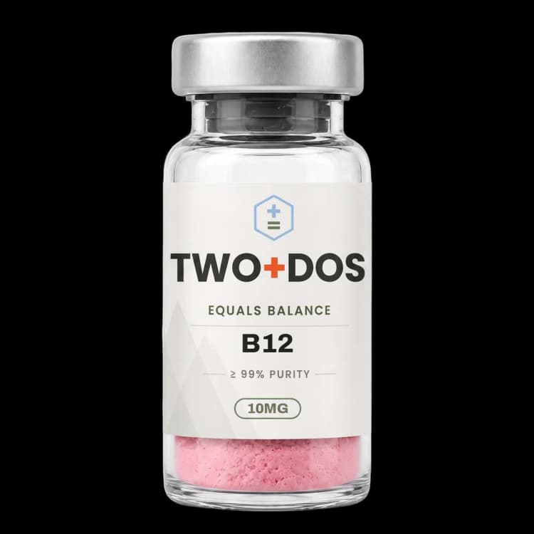
Chemical and physical properties of Vitamin B12 (methylcobalamin)
| Compound class | Cobalt corrinoid coenzyme of the alkylcobalamin subgroup. Not a peptide, and it carries no amino acid sequence |
|---|---|
| Molecular formula | C63H91CoN13O14P, differing from cyanocobalamin by exchange of the upper cyanide ligand for a methyl group |
| Molecular mass | 1344.4 g/mol; exact and monoisotopic mass 1343.5878 Da (PubChem computed) |
| CAS number | 13422-55-4, assigned to mecobalamin |
| PubChem CID | 10898559, listed under the synonyms methylcobalamin and mecobalamin |
| Upper axial ligand | A methyl group bound to cobalt through a covalent metal-carbon sigma bond, one of two such linkages in mammalian biochemistry |
| Lower axial ligand | 5,6-dimethylbenzimidazole, tethered through a ribose-phosphate-aminopropanol loop, coordinating cobalt intramolecularly in the base-on form at neutral pH |
| Cobalt oxidation states | Co(III) in the resting alkylated molecule; Co(II) after homolytic cleavage; Co(I) after heterolytic cleavage, the strongly nucleophilic species used in methyl transfer |
| Ultraviolet-visible maxima | 266, 342 and 522 nm in phosphate buffer at pH 7.0; shifting to 264, 304 and 462 nm in 0.1 N hydrochloric acid as the benzimidazole protonates and detaches |
| Physical description | Dark red crystalline solid, decomposing above 190 °C; sparingly soluble in cold water, more soluble hot |
| Photosensitivity | The cobalt-carbon bond photolyses under visible and ultraviolet light, so identity and purity work is carried out under subdued lighting in amber glassware |
What is methylcobalamin and how is the molecule assembled?
Methylcobalamin belongs to the corrinoid family: a cobalt ion held in a partially reduced tetrapyrrole ring called corrin, carrying a nucleotide loop that folds back to coordinate the metal from below. The upper coordination site holds a methyl group joined to cobalt through a genuine metal-carbon bond.
Corrin differs from porphyrin in two structural details that matter chemically. One of the four methine bridges is missing, so rings A and D are joined directly, and the macrocycle is more reduced and less symmetric than a porphyrin. That lowered symmetry decouples the spectroscopic signature of the trans-axial ligand from that of the methyl group, a point exploited analytically in the X-ray absorption work described further below. Six amide side chains decorate the periphery, and one of them extends into the aminopropanol-phosphate-ribose arm that terminates in 5,6-dimethylbenzimidazole.
Two coordination states are described throughout the literature. In the base-on form the benzimidazole nitrogen occupies the lower axial position; in the base-off form it swings away and is replaced by water, by a protein-supplied histidine, or by nothing at all. Acid drives the free molecule base-off by protonating the benzimidazole, which is visible as a shift of the ultraviolet-visible maxima from 266, 342 and 522 nm at pH 7.0 to 264, 304 and 462 nm in 0.1 N hydrochloric acid. Mammalian enzymes exploit the same switch deliberately, displacing the intramolecular base with a histidine residue.
How does methylcobalamin serve as the methyl carrier of methionine synthase?
Methylcobalamin is the working cofactor of methionine synthase, the cobalamin-dependent enzyme designated MTR in humans and MetH in bacteria. The enzyme moves a methyl group from 5-methyltetrahydrofolate onto homocysteine, producing methionine and tetrahydrofolate, and the cobalt centre serves as the intermediate that carries that methyl group between the two substrates.
The cycle alternates between two oxidation states. Enzyme-bound methylcobalamin transfers its methyl group to the thiolate of homocysteine, leaving cob(I)alamin, a species nucleophilic enough that chemists call it supernucleophilic. Cob(I)alamin then abstracts a methyl group from 5-methyltetrahydrofolate, restoring the methylated Co(III) state. Because Co(I) is easily oxidised, the enzyme drifts into an inactive cob(II)alamin form and must be repaired by reductive methylation using methionine synthase reductase, NADPH and S-adenosylmethionine.
Structural work has caught up with the mechanism only recently. Mendoza and colleagues reported the first full-length structure of a cobalamin-dependent methionine synthase in Nature Communications in 2023, using a thermophilic homologue and capturing cofactor loading in crystallo. Ferreira and colleagues followed in 2026 with cryo-electron microscopy structures of the human enzyme in apo and cobalamin-bound states, showing that the two halves of the apo protein act independently until cofactor binding produces a flexible catalytically competent arrangement, and that methionine synthase reductase engages two distinct sites within the cobalamin-binding half.
Kinetics show how narrow the tolerance is. Guha and Banerjee reported in the Journal of Biological Chemistry in 2025 that the P1173L variant of human MTR retains near-normal specific activity when driven by an artificial reducing system, yet shows a Michaelis constant for S-adenosylmethionine roughly 40-fold higher than wild type and roughly 30-fold lower activity under the physiologically relevant reductase-plus-NADPH system. Pre-steady-state analysis placed electron transfer from the reductase to cob(II)alamin as the rate-limiting step, so the defect lies in reactivation rather than in methyl transfer itself.
How do cells acquire cobalamin and convert it into methylcobalamin?
Methylcobalamin is not taken up directly by mammalian cells. Circulating cobalamin travels bound to transcobalamin, enters through the receptor CD320, and passes through the lysosome before cytosolic enzymes strip its upper ligand and rebuild the methylated form. Each step in that route has been mapped structurally within the past three years.
CD320 turned out to be more complex than the textbook monomer. Guo and colleagues reported in 2026 that the receptor sits at the surface of HEK293 cells as an oligomer of five homomers cross-linked by disulfide bonds, assembled in the endoplasmic reticulum, requiring the transmembrane domain but neither N- nor O-glycosylation for oligomerisation itself. Deleting the low-density-lipoprotein-receptor-like domains abolished both oligomerisation and surface localisation and cut cobalamin uptake. Earlier work from the same group had shown that O-glycans, rather than N-glycans, control trafficking of the receptor to the surface, with the three N-glycosylation sites Asn126, Asn195 and Asn213 dispensable for that step.
Export from the lysosome depends on a transporter-chaperone pair. Liu and colleagues published cryo-electron microscopy structures in Nature Communications in 2026 of the cobalamin exporter ABCD4 in complex with LMBD1, resolved in lumen-open, substrate-bound and cytosol-open states. LMBD1 contributes nine transmembrane helices and a cytosolic domain, both engaging ABCD4, and disrupting those contacts left the exporter unable to reach lysosomes in cell imaging.
In the cytosol the processing enzyme CblC, encoded by MMACHC, removes the upper axial ligand. Esser and colleagues characterised that reaction in Biochemistry in 2025, showing glutathione-driven demethylation of methylcobalamin that yields aquacobalamin together with a stoichiometric equivalent of S-methylglutathione. Replacing the conserved Cys149 with serine or alanine accelerated the observed demethylation rate while uncoupling electron transfer and raising oxidised glutathione output, indicating that the residue tunes the enzyme toward economy rather than speed. Downstream, the chaperone MMADHC delivers the freed cobalamin to methionine synthase, an interaction confirmed by the 2026 cryo-electron microscopy study.
What makes the cobalt-carbon bond of methylcobalamin distinctive?
The cobalt-carbon bond of methylcobalamin is one of only two organometallic linkages known in mammalian biochemistry, the other being the cobalt-adenosyl bond of adenosylcobalamin. Bond strength, photosensitivity and the identity of the trans-axial ligand together decide whether that bond breaks homolytically into radicals or heterolytically into ions.
Gas-phase measurements on the base-off cobinamide analogues place homolytic bond dissociation values at 44.6 plus or minus 0.8 kcal/mol for methylcobinamide against 41.5 plus or minus 1.2 kcal/mol for adenosylcobinamide, the methyl species being the stronger of the pair. Enzymes shift these numbers by remodelling the lower axial position, which is why the trans-axial ligand is studied so closely. Zhao and colleagues applied cobalt K-edge X-ray absorption spectroscopy in 2026 across methylcobalamin, methylcobinamide and the methylated corrinoid iron-sulfur protein of the Wood-Ljungdahl pathway, finding near-identical cobalt-methyl distances yet a systematic rise in pre-edge intensity and a shift to lower transition energy. Their analysis assigned that trend to the trans-axial distance and concluded that the methylated protein retains a weakly bound water rather than being five-coordinate.
Photochemistry provides the second handle. Mukherjee and colleagues reported in Chemical Science in 2026 that anaerobic photolysis of alkylcobalamins in aqueous solution generates Co(I) in nearly quantitative yield, but that adding alkyl halide to that Co(I) does not regenerate the Co(III)-alkyl species quantitatively as its nucleophilicity would predict. A branching pathway intervenes, and both the final cobalt oxidation state and the fate of the organic radical proved solvent-dependent. Computational work published earlier had shown that molecular oxygen redirects methylcobalamin photolysis entirely, with the released methyl radical combining with dioxygen and recombining with the cobalt(II) corrin instead of escaping.
Which microbial systems use the same methyl-cobalt chemistry?
Methylcorrinoid chemistry reaches far beyond methionine synthase. Anaerobic bacteria and archaea run whole metabolisms on methyl-cobalt intermediates, transferring methyl groups from quaternary amines, methoxylated aromatics, halogenated alkanes and carbon monoxide onto corrinoid proteins that are chemically close relatives of methylcobalamin itself.
Picking and colleagues delineated the active site of MtgB, a cobalamin-dependent glycine betaine methyltransferase from Desulfitobacterium hafniense, in the Journal of Biological Chemistry in 2026. Two aromatic residues, Tyr97 and Phe356, engage the quaternary amine through pi-cation contacts while His348 and Arg312 hold the carboxylate, positioning the substrate for nucleophilic attack by the Co(I) centre. Modelling other members of the MttB superfamily placed the quaternary nitrogen of each substrate within an average of 1.8 Å of the others inside the TIM-barrel fold, implying a shared geometry for methyl delivery to cobalt.
Two 2026 papers extended the same logic to different chemistry. Bernhardt and colleagues identified the cdmBCA gene cluster of the acetogen Acetobacterium dehalogenans in Nature Communications as the long-sought corrinoid-dependent chloromethane methyltransferase system, with a crystal structure of CdmB revealing a hydrophobic channel steering haloalkanes toward cobalamin-dependent activation. Bourdin and colleagues characterised glutamine C-methyltransferase in Communications Biology, a cobalamin-dependent radical S-adenosylmethionine enzyme that methylates a glutamine in the active site of methyl-coenzyme M reductase, and showed that hydrogen-atom abstraction and methyl transfer are separate rather than concerted events.
Reliance on cobalamin also creates a vulnerability that shapes microbial communities. Wasson and McRose reported in mBio in 2026 that nitrous oxide inactivates cobalamin-dependent methionine synthase: deleting metE in Pseudomonas aeruginosa, forcing exclusive reliance on the cobalamin-dependent MetH route, sensitised the organism to both exogenous and self-produced nitrous oxide. A substantial fraction of an Arabidopsis thaliana rhizosphere culture collection was found to carry only cobalamin-dependent methionine synthases, and one such isolate was suppressed in co-culture with a nitrous-oxide-producing denitrifier.
What have recent cell-based models shown about methylcobalamin-dependent metabolism?
Cell-based work published since 2025 has used methylcobalamin availability as an experimental variable rather than a fixed background condition. Cancer cell lines, T lymphocyte lines and brain-specific knockout mice each expose a different consequence of interrupting the methylcobalamin-dependent branch of one-carbon metabolism, and the readouts are metabolic rather than descriptive.
Husseiny and Nilsson addressed a decades-old puzzle in Cancer and Metabolism in 2025. Many cancer cell lines fail to grow when methionine in the medium is replaced by homocysteine, despite carrying an intact methionine synthase gene. Culturing such lines with high cobalamin for extended periods produced revertants able to grow on homocysteine, and metabolic flux analysis showed the parental cells never fully engaging methionine synthase. Expressing a cobalamin-independent methionine synthase restored growth and normalised the ratio of S-adenosylmethionine to S-adenosylhomocysteine, whereas overexpressing the cobalamin-dependent human enzyme did nothing, locating the limitation in cofactor supply rather than in enzyme abundance.
A separate 2026 report placed methylcobalamin in an unrelated role. Wang and colleagues, working in Virologica Sinica, characterised gasdermin-E-dependent pyroptosis triggered by enterovirus A71 in Jurkat and EL-4 T cell lines and in primary mouse CD3-positive T cells, driven through caspase-3 rather than through gasdermin D. Methylcobalamin, applied as a gasdermin E inhibitor, limited the resulting T-cell loss in culture and raised survival in an infected newborn mouse model to 80 percent. The observation is notable because the molecule acts there as a direct protein inhibitor rather than as an enzyme cofactor.
Summary of published research
Findings below are reported as published by the cited authors, in the model systems they used. They describe laboratory research, and none of them characterize use in humans.
Ferreira DSM, McLennan K, Diamond C, et al. Structural insights into cobalamin loading and reactivation of human methionine synthase. Nature Communications (2026)
- Model system
- Recombinant human methionine synthase; cryo-electron microscopy and AlphaFold-guided interaction studies
- Conditions
- Apo and cobalamin-bound states of full-length MTR, with MMADHC and methionine synthase reductase
- Reported finding
- Apo MTR adopted a conformation in which the catalytic and cobalamin-binding halves acted independently, and cofactor binding produced a flexible catalytically competent state. MMADHC engaged the cobalamin-binding half of the apo enzyme, and the reductase was found to contact two distinct sites within that half.
Guha A, Banerjee R. The common homocystinuria-associated P1173L variant of human methionine synthase impairs reductive methylation. Journal of Biological Chemistry (2025)
- Model system
- Purified recombinant human MTR co-expressed with the MMADHC chaperone; steady-state, pre-steady-state and EPR analysis
- Conditions
- Artificial reducing system compared against methionine synthase reductase with NADPH and S-adenosylmethionine
- Reported finding
- The variant showed a Michaelis constant for S-adenosylmethionine roughly 40-fold above wild type and roughly 30-fold lower activity with the reductase-based system, while complex formation with the reductase was unaffected. Electron transfer to cob(II)alamin became rate-limiting.
Guo W, Qiu R, Zhao X, et al. Transcobalamin receptor CD320, responsible for vitamin B12 cellular uptake, is present on the cell surface as a homo-oligomer. Journal of Biological Chemistry (2026)
- Model system
- Transfected human embryonic kidney 293 cells; glycosidase digestion, western blotting and uptake assays
- Conditions
- Wild-type CD320, domain-deletion mutants and the delta-E88 variant
- Reported finding
- CD320 reached the cell surface as a disulfide-linked assembly of five homomers formed in the endoplasmic reticulum. Removing the LDL-receptor-like domains abolished oligomerisation, surface localisation and cobalamin uptake, and the delta-E88 variant slowed exit from the endoplasmic reticulum.
Du C, Guo W, Wang M, et al. O-glycosylation is essential for cell surface expression of the transcobalamin receptor CD320. Journal of Biological Chemistry (2024)
- Model system
- Transfected HEK293 and HepG2 hepatoma cells; site-directed mutagenesis, flow cytometry and lectin binding
- Conditions
- N-glycosylation site substitutions at Asn126, Asn195 and Asn213 versus chemical inhibition of O-glycosylation
- Reported finding
- Glycosylation and sialylation produced an approximately 70 kDa species forming a high-molecular-weight surface complex. Abolishing all three N-glycosylation sites left trafficking intact, whereas blocking O-glycosylation reduced surface CD320 and lowered cobalamin uptake.
Liu Q, Li X, Wu Y, et al. Structural basis for LMBD1-dependent trafficking and cobalamin export of ABCD4. Nature Communications (2026)
- Model system
- Purified human ABCD4-LMBD1 complex; cryo-electron microscopy plus cell imaging
- Conditions
- Lumen-open, substrate-bound and cytosol-open conformational states
- Reported finding
- LMBD1 was resolved with nine transmembrane helices and a cytosolic domain, both contacting the lysosomal cobalamin exporter ABCD4. Disrupting those contacts impaired delivery of ABCD4 to lysosomes, and the structures defined cobalamin recognition across the transport cycle.
Esser AJ, Sastre S, Dinh TJ, et al. A Noncatalytic Cysteine Residue Modulates Cobalamin Reactivity in the Human B12 Processing Enzyme CblC. Biochemistry (2025)
- Model system
- Purified recombinant human CblC (MMACHC) with Cys149Ser and Cys149Ala substitutions; cell-free kinetics
- Conditions
- Glutathione-driven dealkylation of methylcobalamin, aerobic and long-incubation conditions
- Reported finding
- Demethylation of methylcobalamin yielded aquacobalamin and a stoichiometric equivalent of S-methylglutathione. Both Cys149 substitutions accelerated the observed dealkylation rate while significantly uncoupling electron transfer, and allowed conversion of aquacobalamin to cob(II)alamin under oxygen.
Mukherjee A, Wu SY, Cooper DJ, et al. Shining light on the mechanism of photochemical alkene formation in vitamin B12. Chemical Science (2026)
- Model system
- Cell-free alkylcobalamin solutions; ultrafast and single-turnover time-resolved spectroscopy
- Conditions
- Anaerobic aqueous photolysis, then alkyl halide addition across different solvents
- Reported finding
- Anaerobic photolysis generated Co(I) in nearly quantitative yield, yet alkyl halide addition did not regenerate the Co(III)-alkyl species quantitatively, revealing a branching pathway. Final cobalt oxidation state and organic radical fate were both solvent-dependent.
Zhao K, Abernathy MJ, Griffith C, et al. Investigating weak axial ligation in corrinoids by X-ray absorption spectroscopy: Implications for corrinoid iron-sulfur protein. Journal of Inorganic Biochemistry (2026)
- Model system
- Cell-free corrinoid series: methylcobalamin, methylcobinamide and methylated corrinoid iron-sulfur protein
- Conditions
- Cobalt K-edge X-ray absorption spectroscopy with DFT, TD-DFT and QM/MM analysis
- Reported finding
- Cobalt-methyl distances were similar across the series, while the pre-edge feature rose in intensity and shifted to lower transition energy. The trend was assigned to the trans-axial distance, supporting a weakly bound water on the methylated protein rather than a five-coordinate centre.
Picking J, Li Y, Ticak T, et al. Delineation of the active site of MtgB, a cobalamin-dependent glycine betaine methyltransferase. Journal of Biological Chemistry (2026)
- Model system
- Desulfitobacterium hafniense MtgB; site-directed substitution plus X-ray crystallography of the substrate-bound enzyme
- Conditions
- Glycine betaine to cob(I)alamin methyl transfer assays
- Reported finding
- Tyr97 and Phe356 contacted the quaternary amine through pi-cation interactions while His348 and Arg312 held the carboxylate, each residue proving necessary for catalysis. Modelling across the MttB superfamily placed substrate nitrogens within an average of 1.8 Å inside the TIM barrel.
Wasson PA, McRose DL. Nitrous oxide produced by denitrifying pseudomonads inhibits the growth of rhizosphere bacteria by inactivating the cobalamin-dependent methionine synthase. mBio (2026)
- Model system
- Pseudomonas aeruginosa metE deletion mutants and an Arabidopsis thaliana rhizosphere culture collection
- Conditions
- Exogenous and self-generated nitrous oxide; co-culture with a nitrous-oxide-producing denitrifier
- Reported finding
- Forcing reliance on cobalamin-dependent MetH sensitised the organism to nitrous oxide. A substantial share of the rhizosphere collection carried only cobalamin-dependent methionine synthases and proved sensitive, and one isolate was suppressed in co-culture.
Husseiny MMAE, Nilsson R. The role of B12 deficiency and methionine synthase in methionine-dependent cancer cells. Cancer & Metabolism (2025)
- Model system
- Cancer cell lines cultured with homocysteine in place of methionine; single-cell RNA sequencing and metabolic flux analysis
- Conditions
- Long-term culture with high cobalamin; rescue by a cobalamin-independent methionine synthase
- Reported finding
- Methionine-dependent lines did not fully engage methionine synthase in homocysteine medium. High cobalamin generated revertants able to grow, and expressing a cobalamin-independent enzyme restored growth and normalised the S-adenosylmethionine to S-adenosylhomocysteine ratio.
Wang C, Guo B, Li Y, et al. EV-A71 induces GSDME-dependent T-cell pyroptosis: A novel mechanism and MeCbl therapeutic potential. Virologica Sinica (2026)
- Model system
- Jurkat and EL-4 T cell lines, primary mouse CD3-positive T cells, and newborn mice
- Conditions
- Enterovirus A71 infection with genetic ablation and inhibitor controls
- Reported finding
- Infection triggered gasdermin-E-dependent pyroptosis through caspase-3 rather than gasdermin D. Methylcobalamin, acting as a gasdermin E inhibitor, limited T-cell loss in culture and raised survival in the infected newborn mouse model to 80 percent.
What laboratory handling information is published?
Methylcobalamin is supplied as a dark red crystalline solid that decomposes above 190 °C rather than melting cleanly. Published property data describe it as sparingly soluble in cold water and more soluble in hot, and material is normally kept sealed against moisture in a freezer at or below −20 °C. The corrin ring accounts for the colour: aqueous solutions are visibly red-pink even at low micromolar concentration.
Light is the dominant stability variable, and it is a chemical rather than a nuisance problem. The cobalt-carbon bond absorbs in the visible region and cleaves photolytically, so ordinary bench lighting slowly converts methylcobalamin into cob(II)alamin, aquacobalamin and related species. Published spectroscopic work shows the outcome depends on whether oxygen is present: under anaerobic conditions the cobalt centre can be driven nearly quantitatively to Co(I), while under air the released methyl radical combines with dioxygen and recombines with the cobalt(II) corrin. Standard practice is amber glassware or foil-wrapped vessels, subdued room lighting, and solutions prepared shortly before measurement.
Identity is confirmed spectroscopically before anything else. Ultraviolet-visible scanning in phosphate buffer at pH 7.0 should reproduce maxima near 266, 342 and 522 nm, and acidifying the same sample to 0.1 N hydrochloric acid should shift them to approximately 264, 304 and 462 nm as the benzimidazole protonates and the molecule converts to the base-off form. That pH-dependent shift distinguishes an alkylcobalamin from cyanocobalamin, whose spectrum is dominated by a band near 361 nm. Mass spectrometry against a monoisotopic mass of 1343.5878 Da and reversed-phase HPLC purity on the lot certificate of analysis complete the identity package.
Two contamination questions recur in method sections. First, photodegradation products are themselves cobalamins and remain red, so visual inspection tells nothing; chromatography is required. Second, cyanide traces during synthesis or workup convert alkylcobalamins to cyanocobalamin, which is why suppliers report the four cobalamin forms separately rather than as total corrinoid.
This material is offered for laboratory research use only. It is not a drug, not a food, and not intended for human or veterinary use.
Frequently asked research questions
Is methylcobalamin a peptide?
No. Methylcobalamin is a cobalt coordination complex built on a corrin macrocycle, with a molecular formula of C63H91CoN13O14P and no amino acid sequence at all. It is grouped with research peptides only because laboratories order it alongside them; chemically it belongs with the metallocofactors.
How does methylcobalamin differ from cyanocobalamin and adenosylcobalamin?
All three share the same corrin-cobalt core and differ only in the upper axial ligand. Cyanocobalamin carries cyanide, which makes it photostable and cheap to manufacture, so it dominates industrial production. Methylcobalamin carries a methyl group and adenosylcobalamin a 5-deoxyadenosyl group; both are alkylcobalamins, both are photolabile, and both are the forms enzymes actually use.
What do base-on and base-off mean for methylcobalamin?
Base-on describes the state in which the tethered 5,6-dimethylbenzimidazole coordinates cobalt from below, which is how the free molecule sits at neutral pH. Base-off describes displacement of that group by acid, by water, or by a protein-supplied histidine. The switch changes reactivity: X-ray absorption work published in 2026 attributed differences in cobalt pre-edge spectra across methylcobalamin, methylcobinamide and a methylated corrinoid protein to exactly this trans-axial position.
Which enzymes in mammalian cells depend on methylcobalamin?
One, in the strict sense. Methionine synthase is the only mammalian enzyme that uses methylcobalamin, transferring a methyl group from 5-methyltetrahydrofolate to homocysteine. A second mammalian enzyme, methylmalonyl-CoA mutase, uses adenosylcobalamin instead. Everything else in the cobalamin literature concerns the transport, processing and chaperone machinery that supplies those two.
What model systems dominate the recent methylcobalamin literature?
Four recur across the citations above: cell-free spectroscopy and enzyme kinetics on purified corrinoids and methyltransferases; cryo-electron microscopy of human transport and cofactor-loading complexes; transfected HEK293 and HepG2 cells for receptor trafficking and uptake; and bacterial genetics in Pseudomonas, Desulfitobacterium and Acetobacterium for methyl-transfer chemistry.
Vitamin B12 (methylcobalamin) at TWO+DOS
TWO+DOS supplies Vitamin B12 (methylcobalamin) as a research-use-only compound, third-party tested, with certificates of analysis emailed immediately on request.
View the B12 (10mg)listing →Related research overviews
References
- PubChem CID 10898559: Methylcobalamin compound summary
- Structure of full-length cobalamin-dependent methionine synthase and cofactor loading captured in crystallo (Nature Communications 2023)
- Structural insights into cobalamin loading and reactivation of human methionine synthase (Nature Communications 2026)
- The common homocystinuria-associated P1173L variant of human methionine synthase impairs reductive methylation (Journal of Biological Chemistry 2025)
- Transcobalamin receptor CD320 is present on the cell surface as a homo-oligomer (Journal of Biological Chemistry 2026)
- O-glycosylation is essential for cell surface expression of the transcobalamin receptor CD320 (Journal of Biological Chemistry 2024)
- Structural basis for LMBD1-dependent trafficking and cobalamin export of ABCD4 (Nature Communications 2026)
- A Noncatalytic Cysteine Residue Modulates Cobalamin Reactivity in the Human B12 Processing Enzyme CblC (Biochemistry 2025)
- Shining light on the mechanism of photochemical alkene formation in vitamin B12 (Chemical Science 2026)
- Investigating weak axial ligation in corrinoids by X-ray absorption spectroscopy (Journal of Inorganic Biochemistry 2026)
- Delineation of the active site of MtgB, a cobalamin-dependent glycine betaine methyltransferase (Journal of Biological Chemistry 2026)
- The role of B12 deficiency and methionine synthase in methionine-dependent cancer cells (Cancer & Metabolism 2025)
For research use only. Not for human or veterinary use. Not for diagnostic or therapeutic use.