
LIPO-C (Methionine, Inositol, Choline, Cyanocobalamin): Research Overview
By the TWO+DOS Research Team · Published 2026-08-13
For research use only. Not for human or veterinary use. Not for diagnostic or therapeutic use.
LIPO-C is a four-component laboratory blend of L-methionine, myo-inositol, choline and cyanocobalamin. The four molecules span three chemical classes: a sulfur-containing amino acid at 149.21 g/mol, a meso cyclitol at 180.16 g/mol, a quaternary ammonium cation, and a 1355.4 g/mol cobalt corrinoid carrying an axial cyanide ligand.
What follows is restricted to molecular-level material: identifiers and structural chemistry, the carrier proteins and enzymes each component engages, the measured affinities and structures published for those proteins, and the analytical and stability behaviour that governs how a mixture of this composition is characterised in a laboratory. Outcome literature is outside the scope of this page.
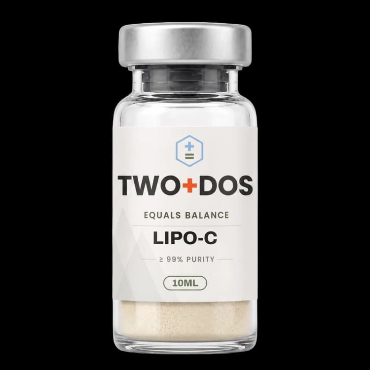
Chemical and physical properties of LIPO-C (methionine, inositol, choline, cyanocobalamin)
| Composition class | Four small molecules spanning three chemical families: a sulfur-bearing amino acid, a cyclitol polyol, a quaternary ammonium cation, and a cobalt corrinoid coordination complex |
|---|---|
| L-Methionine | C5H11NO2S, 149.21 g/mol, CAS 63-68-3, PubChem CID 6137; one stereocentre, L configuration |
| myo-Inositol | C6H12O6, 180.16 g/mol, CAS 87-89-8, PubChem CID 892; IUPAC name cyclohexane-1,2,3,4,5,6-hexol |
| Choline chloride | C5H14ClNO, 139.62 g/mol, CAS 67-48-1, PubChem CID 6209; the free choline cation is C5H14NO+ at 104.17 g/mol (CID 305) |
| Cyanocobalamin | C63H88CoN14O14P, 1355.4 g/mol, CAS 68-19-9, PubChem CID 166596686; cobalt(III) held in a corrin macrocycle with axial cyanide and 5,6-dimethylbenzimidazole ligands |
| Aqueous solubility | L-methionine 56.6 g/L at 25 °C; myo-inositol roughly 14 g per 100 mL at 25 °C, rising above 27 g per 100 mL at 50 °C; choline chloride deliquescent and miscible in all proportions |
| Ionisation | L-methionine pKa1 2.28 and pKa2 9.21, zwitterionic across the neutral range; choline carries a permanent formal positive charge that no pH change removes |
| Polarity | L-methionine log Kow -1.87; all four components are strongly hydrophilic, so none partitions into a lipid bilayer without carrier assistance |
| Stereochemistry | myo-Inositol carries an internal mirror plane (five equatorial hydroxyls, one axial) and is therefore meso and optically inactive despite six substituted carbons |
| Chromophore | Only cyanocobalamin absorbs usefully in the visible and near-ultraviolet, with maxima near 278, 361 and 550 nm; the 550 nm band produces the deep red colour |
| Photolability | The cobalt-carbon axis of the corrinoid is light-sensitive, so cobalamin work is performed under shielded light; the other three components are photochemically inert |
Why are these four molecules grouped in one preparation?
LIPO-C groups four molecules that converge on a single biochemical node: the transfer of methyl groups. Methionine supplies the methyl donor S-adenosylmethionine, choline supplies methyl groups after oxidation to betaine, cyanocobalamin supplies the cofactor of methionine synthase, and myo-inositol sits adjacent as the headgroup precursor of phosphatidylinositol.
Homocysteine is the junction where three of the four meet. Two independent enzymes remethylate it. Methionine synthase (MTR) uses a cobalamin cofactor and takes its methyl group from 5-methyltetrahydrofolate. Betaine-homocysteine S-methyltransferase (BHMT) bypasses cobalamin entirely, taking its methyl group from betaine, the oxidation product of choline. BHMT is a zinc metalloenzyme; reported Michaelis constants are near 2.2 mM for betaine and 4 μM for L-homocysteine, and the enzyme is activated by potassium with an apparent constant near 100 μM. Choline therefore feeds methionine chemistry through a route that has no cobalamin dependence at all, which is the structural logic behind pairing the two in one preparation.
Choline reaches betaine through two oxidation steps. Choline dehydrogenase (CHDH), a flavin enzyme of the inner mitochondrial membrane, oxidises choline to betaine aldehyde, and an aldehyde dehydrogenase (ALDH7A1 and related isoforms) completes the conversion to betaine. Only a fraction of cellular choline takes this route; the remainder is phosphorylated by choline kinase and committed to phosphatidylcholine synthesis, a branch point discussed below.
myo-Inositol is the outlier in metabolic terms. The cyclitol carries no transferable methyl group and participates in no one-carbon reaction. Its connection to the other three is compositional rather than catalytic: phosphatidylinositol and phosphatidylcholine are both glycerophospholipids assembled from CDP-activated intermediates, so inositol and choline supply alternative headgroups drawn into overlapping lipid pools.
Which transporters carry choline across the plasma membrane?
Choline, the quaternary ammonium component of LIPO-C, holds a permanent positive charge and cannot traverse a lipid bilayer unaided. Three carrier families have been proposed to move it: the high-affinity transporter CHT1 (SLC5A7), the choline transporter-like SLC44 proteins, and the SLC49 proteins FLVCR1 and FLVCR2, which were reassigned from haem transport to choline transport in 2024.
The 2024 reassignment rests on structural and kinetic data. Cryo-electron microscopy resolved FLVCR1 and FLVCR2 in inward-facing states at 2.9 Å, an outward-facing FLVCR2 at 3.1 Å, and substrate-bound states at 2.6 and 2.8 Å. Transport measurements returned choline Michaelis constants of 47.4 μM for FLVCR1 and 64.0 μM for FLVCR2, with ethanolamine handled more tightly by FLVCR1 at 8 μM. Selectivity traces to a conserved aromatic pair, Trp125 and Tyr349 in FLVCR1 and Trp102 and Tyr325 in FLVCR2, whose π systems engage the substrate ammonium group through cation-pi contacts rather than through hydrogen bonding.
A second 2024 structural study reached the same assignment from the lipid side, describing FLVCR1 as the entry point that delivers choline and ethanolamine to their respective kinases at the head of the Kennedy pathway. Structure-guided mutagenesis in that work separated the two activities: certain residues proved indispensable for ethanolamine movement while leaving choline movement intact, which functionally uncouples the two branches of phospholipid headgroup supply.
The status of the SLC44 family has narrowed as a result. A 2026 comparison of FLVCR and CTL proteins across eukaryotes supported FLVCR proteins as genuine choline transporters and suggested a non-transport role for CTL proteins, which had previously been credited with intermediate-affinity choline uptake in the range of roughly 5 to 50 μM. CHT1 remains the highest-affinity route, with constants reported between about 0.5 and 5 μM, and a 2026 cryo-electron microscopy study of a bacterial sodium-dependent choline transporter placed it in the LeuT fold with conserved sodium coordination, arguing for a translocation mechanism shared between the bacterial protein and human CHT1.
Directionality has also been mapped in an epithelial model. Work published in 2025 using Caco-2 monolayers and rat small intestine assigned FLVCR2 to apical uptake and FLVCR1 to basolateral efflux, with knockdown reducing cellular accumulation and identified sequence variants lowering activity. The two carriers act in series rather than in parallel, which is why a single-carrier description of choline movement across an epithelium fails.
How does cyanocobalamin become a catalytically competent cofactor?
Cyanocobalamin, the corrinoid in LIPO-C, is catalytically inert as supplied. The cobalt(III) centre sits in a corrin macrocycle with 5,6-dimethylbenzimidazole below the ring and cyanide occupying the upper axial position, and that cyanide must be removed before the metal can carry a methyl group. The chaperone MMACHC, the product of the cblC locus, performs the removal.
MMACHC carries an N-terminal flavodoxin-like nitroreductase domain and catalyses reductive decyanation using FMN or FAD together with NADPH, releasing cyanide and generating the supernucleophilic cob(I)alamin. Reducing equivalents are furnished in cells by cytosolic diflavin oxidoreductases, methionine synthase reductase and novel reductase 1. The same protein dealkylates alkylcobalamins by a glutathione-dependent route, so one chaperone strips both cyanide and alkyl groups from incoming corrinoids and hands over a common cobalt(I) product.
Downstream loading has been resolved structurally. A 2026 cryo-electron microscopy study of full-length human methionine synthase determined the enzyme in apo and cobalamin-bound states, showing that the catalytic N-half and the cobalamin-binding C-half behave as independent modules in the apo form, with the C-half positioned for cofactor capture. Cofactor binding and activation drive the assembly into a flexible catalytically competent arrangement. Interaction mapping placed MMADHC on the C-half of the apo enzyme, consistent with a role in cofactor delivery, and located methionine synthase reductase at two separate C-half sites.
Cyanide stoichiometry is worth stating plainly because it is often misread. One cyanide ion accompanies each corrinoid molecule, so a 1355.4 g/mol species carries 26.02 g/mol of cyanide, about 1.9 percent by mass. The ligand is what makes this the most photochemically robust and crystallographically tractable of the common cobalamins, and it is also the reason the compound requires enzymatic processing before any methyl chemistry can begin.
What enzymes act on methionine once it enters a cell?
Methionine in LIPO-C is the L-enantiomer, CAS 63-68-3, a 149.21 g/mol amino acid whose thioether side chain gives it a log Kow of -1.87 and pKa values of 2.28 and 9.21. Intracellular processing begins with methionine adenosyltransferase, which condenses the amino acid with ATP to build the sulfonium methyl donor S-adenosylmethionine.
The somatic isoform MAT2A follows a strictly ordered kinetic mechanism in which ATP binds before L-methionine and S-adenosylmethionine leaves before phosphate and pyrophosphate depart in random order. That ordering has been exploited in medicinal chemistry: allosteric MAT2A inhibitors are under clinical evaluation in tumours lacking methylthioadenosine phosphorylase, and a 2026 pyridazinone series reported a lead compound that lowered cellular S-adenosylmethionine and symmetric dimethylarginine in MTAP-deleted models. MAT2A shares roughly 84 percent sequence homology with the liver isoform MAT1A and the two superimpose to below 1 Å, which is why selective inhibition has depended on allosteric rather than substrate-competitive chemistry.
S-Adenosylmethionine donates its methyl group to nucleic acids, proteins and small-molecule acceptors, and every such reaction yields S-adenosylhomocysteine, which adenosylhomocysteine hydrolase cleaves to adenosine and homocysteine. The ratio of the two adenosyl species is the standard laboratory index of methylation capacity, and it is the quantity that links methionine supply back to the cobalamin-dependent and betaine-dependent remethylation routes described earlier.
The thioether sulfur is also the most oxidisable group in the blend. Oxidation converts methionine to methionine sulfoxide, generating a new stereocentre at sulfur, and two enzyme families reverse it with opposite specificity: MsrA reduces the S epimer and MsrB the R epimer, both through a sulfenic-acid intermediate resolved by thioredoxin. In an analytical context this matters because methionine sulfoxide is the expected degradation marker for the amino acid component, and a purity record that reports only a main-peak percentage will miss it.
What is established about myo-inositol structure and transport?
myo-Inositol, the cyclitol in LIPO-C, is cyclohexane-1,2,3,4,5,6-hexol at 180.16 g/mol, CAS 87-89-8, PubChem CID 892. Five hydroxyl groups occupy equatorial positions and one is axial. That arrangement places an internal mirror plane through the molecule, making it meso and optically inactive, and it melts in the region of 220 to 227 °C as a hygroscopic crystalline solid.
The meso symmetry creates a specific synthetic problem: preparing a single enantiomer of any O-substituted derivative requires breaking that symmetry. A 2025 review of desymmetrisation surveyed the available approaches, covering classical resolution through separable diastereomers, chiral pool routes, enzyme catalysis, asymmetric catalysis, and preferential crystallisation from racemic conglomerates. The same review notes that laboratory synthesis of enantiomeric inositol derivatives was what made the phosphoinositide signalling field tractable in the first place.
Three mammalian carriers move myo-inositol. SMIT1 (SLC5A3) and SMIT2 (SLC5A11) are sodium-coupled; HMIT (SLC2A13) is proton-coupled. Transport stoichiometry was resolved directly in oocyte work that established two sodium ions per myo-inositol for SMIT2 and a one-to-one proton coupling for HMIT, settling a question that cooperativity data alone had left open. Reported Michaelis constants for SMIT2 sit near 120 μM for myo-inositol and 111 μM for D-chiro-inositol, so the transporter does not sharply discriminate between the two ring stereoisomers.
Export has been characterised much more recently. A 2026 study identified Pho84 in yeast and GLUT2 in mammalian cells as inositol exporters whose activity is governed by inositol pyrophosphates, describing a conserved circuit that couples phosphate signalling to inositol balance. Efflux capacity had previously been the least defined arm of cellular inositol handling.
Inside the cell the cyclitol is consumed almost entirely as a lipid headgroup. Phosphatidylinositol carries it into the plasma membrane, where sequential kinases build phosphatidylinositol 4,5-bisphosphate, and phospholipase C cleavage releases inositol 1,4,5-trisphosphate. Dephosphorylation returns the ring to free inositol through inositol monophosphatase, a magnesium-dependent homodimer of roughly 30 kDa subunits that is inhibited by millimolar lithium; crystallography attributes the inhibition to lithium occupying the second metal site and displacing a catalytic water molecule.
How is a four-component blend characterised analytically?
LIPO-C poses an unusual analytical problem because its four components differ by nearly an order of magnitude in mass and share almost no detection chemistry. Cyanocobalamin absorbs strongly in the visible region, methionine only in the far ultraviolet below about 220 nm, and myo-inositol and choline carry no useful chromophore whatever. No single detector reads all four well.
The corrinoid is the easiest of the four. Its spectrum shows maxima near 278, 361 and 550 nm, and pharmacopoeial identity relies on absorbance ratios rather than a single wavelength, with A361/A278 specified between 1.70 and 1.90 and A361/A550 between 3.15 and 3.40. Because degradation shifts the corrin spectrum rather than abolishing it, those ratios detect conversion that a single-wavelength reading would miss.
Mass spectrometry covers the components that ultraviolet detection cannot. A 2025 LC-MS/MS method separated three cobalamin forms in eight minutes and selected product-ion channels containing fragments unique to active corrinoids, so that inactive analogues could not inflate the result. Hydrophilic interaction chromatography is the usual retention mode for the small polar components, since none is retained on reversed-phase silica without ion pairing; commonly monitored positive-mode transitions are m/z 104 to 60 for choline, 118 to 58 for betaine and 150 to 104 for methionine.
myo-Inositol remains the hardest analyte. Lacking both a chromophore and a readily ionisable group, it is typically measured by refractive index, evaporative light scattering or charged aerosol detection, or by derivatisation before gas chromatography. Published LC-MS/MS assays for the cyclitol report detection limits in the tens of nanograms per millilitre with linear ranges spanning roughly 0.1 to 100 μg/mL, so quantifying it alongside a corrinoid present at a thousandth of its mass usually requires two separate injections rather than one method.
Summary of published research
Findings below are reported as published by the cited authors, in the model systems they used. They describe laboratory research, and none of them characterize use in humans.
Ri K, Weng TH, Claveras Cabezudo A, et al. Molecular mechanism of choline and ethanolamine transport in humans. Nature (2024)
- Model system
- Purified human FLVCR1 and FLVCR2; cryo-electron microscopy with transport assays in a reconstituted system
- Conditions
- Inward-facing maps at 2.9 Å, outward-facing FLVCR2 at 3.1 Å, choline-bound states at 2.6 and 2.8 Å; graded substrate concentrations for kinetics
- Reported finding
- The study measured choline Michaelis constants of 47.4 μM for FLVCR1 and 64.0 μM for FLVCR2 and ethanolamine constants of 8 μM and 41.5 μM respectively, and identified Trp125 with Tyr349 in FLVCR1 and Trp102 with Tyr325 in FLVCR2 as the conserved aromatic pair engaging the substrate ammonium group through cation-pi contacts.
Son Y, Kenny TC, Khan A, Birsoy K, Hite RK. Structural basis of lipid head group entry to the Kennedy pathway by FLVCR1. Nature (2024)
- Model system
- Human FLVCR1 in a lipid environment; cryo-electron microscopy with structure-guided mutagenesis
- Conditions
- Structures determined in the presence of choline and of ethanolamine
- Reported finding
- The work located choline and ethanolamine at a shared site built from aromatic and polar residues, showed that the transporter contacts the quaternary amine of one substrate differently from the primary amine of the other, and identified mutations that abolished ethanolamine movement while leaving choline movement intact.
Nel L, Driller JH, Driller R, Frain KM, Pedersen BP. Structural and biochemical comparison of the FLVCR and CTL membrane protein families in eukaryotes. Life Science Alliance (2026)
- Model system
- Recombinant eukaryotic FLVCR (SLC49) and CTL (SLC44) proteins; comparative structural and biochemical analysis
- Conditions
- Side-by-side expression and substrate testing of both families under matched conditions
- Reported finding
- The comparison supported FLVCR proteins as bona fide choline transporters and reported that CTL proteins did not move choline under the conditions tested, pointing to a non-transport function for the SLC44 family.
Vilchez-Garcia J, Martínez-Jiménez A, Jiang H, et al. Structural insights into a conserved mechanism of choline translocation through CHT. Science Advances (2026)
- Model system
- Bacterial sodium-dependent choline transporter homologous to human CHT1; cryo-electron microscopy with computational analysis
- Conditions
- Structures captured in distinct conformational states with sodium present
- Reported finding
- The structures placed the transporter in the LeuT fold with conserved sodium coordination and supported a translocation mechanism shared between the bacterial protein and the human high-affinity choline transporter CHT1.
Yasujima T, Namba C, Azuma Y, et al. The role of FLVCR1 and FLVCR2 in choline transport in the Caco-2 intestinal epithelial cell model and rat small intestine. Biochimica et Biophysica Acta Molecular Basis of Disease (2025)
- Model system
- Caco-2 monolayers and rat small intestine
- Conditions
- Polarised uptake and efflux measurements with transporter knockdown and variant expression
- Reported finding
- The study assigned apical uptake to FLVCR2 and basolateral efflux to FLVCR1, reported reduced cellular accumulation after knockdown, and characterised sequence variants that lowered transport activity.
Kim J, Gherasim C, Banerjee R. Decyanation of vitamin B12 by a trafficking chaperone. Proceedings of the National Academy of Sciences (2008)
- Model system
- Recombinant human MMACHC (CblC) protein; cell-free spectroscopic and kinetic assay
- Conditions
- Flavin cofactor FMN or FAD with NADPH as the reductant, anaerobic and aerobic comparisons
- Reported finding
- The study established that MMACHC catalyses reductive removal of the axial cyanide ligand from cyanocobalamin, releasing cob(I)alamin, and that flavin plus NADPH are required for the reaction.
Ferreira DSM, McLennan K, Diamond C, et al. Structural insights into cobalamin loading and reactivation of human methionine synthase. Nature Communications (2026)
- Model system
- Recombinant full-length human methionine synthase; cryo-electron microscopy with interaction mapping
- Conditions
- Apo and cobalamin-bound preparations, with AlphaFold-guided analysis of MMADHC and MTRR binding
- Reported finding
- The structures showed the catalytic and cofactor-binding halves acting independently in the apo enzyme, with cofactor capture and activation producing a flexible competent arrangement; MMADHC was placed on the C-half of the apo enzyme and MTRR at two distinct C-half sites.
Bourgeois F, Coady MJ, Lapointe JY. Determination of transport stoichiometry for two cation-coupled myo-inositol cotransporters: SMIT2 and HMIT. Journal of Physiology (2005)
- Model system
- Xenopus laevis oocytes expressing SMIT2 (SLC5A11) or HMIT (SLC2A13)
- Conditions
- Voltage clamp paired with radiolabelled myo-inositol flux measurement
- Reported finding
- The measurements resolved a two-sodium-per-substrate stoichiometry for SMIT2 and one-to-one proton coupling for HMIT, replacing inferences that had rested only on cooperativity in cation activation.
Su XB, Zhang X, Fedeli V, et al. Pho84/GLUT2 are cellular myo-inositol exporters regulated by inositol pyrophosphates. The EMBO Journal (2026)
- Model system
- Saccharomyces cerevisiae genetics with mammalian cell validation
- Conditions
- Manipulation of inositol pyrophosphate levels with tracking of inositol efflux
- Reported finding
- The study identified Pho84 as an inositol exporter in yeast and GLUT2 as its mammalian counterpart, and described an evolutionarily conserved circuit in which inositol pyrophosphates couple phosphate signalling to cellular inositol balance.
Patil NT, Patil MT, Gonnade RG, Shashidhar MS. Desymmetrization of myo-inositol. Carbohydrate Research (2025)
- Model system
- Synthetic organic chemistry review with results from the authors' laboratory
- Conditions
- Comparison of resolution, chiral pool, enzymatic, asymmetric-catalytic and crystallisation strategies
- Reported finding
- The review catalogued the routes by which the mirror symmetry of the meso cyclitol is broken to give single-enantiomer O-substituted derivatives, including preferential crystallisation of enantiomers from racemic conglomerates.
Zhang Y, Wang P, Xie Z, et al. Design and Discovery of Pyridazinone-Based MAT2A Inhibitors Targeting MTAP-Deficient Cancers. Journal of Medicinal Chemistry (2026)
- Model system
- Enzyme assays with MTAP-deleted tumour cell lines and murine xenografts
- Conditions
- Structure-activity series around a pyridazinone scaffold
- Reported finding
- The series produced a lead compound that inhibited MAT2A and lowered cellular S-adenosylmethionine and symmetric dimethylarginine in MTAP-deficient models, linking blockade of the adenosyltransferase step directly to methyl-donor supply.
Fan Z, Li Y, Fan X, Wang P, Yang R, Xie C. Simultaneous Determination of Three Active Forms of Vitamin B12 In Situ Produced During Fermentation by LC-MS/MS. Foods (2025)
- Model system
- Analytical method development on fermented rice bran and Propionibacterium freudenreichii cell mass
- Conditions
- Eight-minute liquid chromatography gradient with tandem mass spectrometric product-ion selection
- Reported finding
- The method resolved adenosyl-, methyl- and hydroxocobalamin using ion channels containing fragments unique to active corrinoids, and reported that light exposure during extraction and clean-up reduced measured corrinoid content by roughly 30 percent.
What laboratory handling information is published?
LIPO-C handling records are dominated by the corrinoid, which is by a wide margin the least robust of the four components. The cobalt-carbon axis of cyanocobalamin is photolabile, and published photolysis work describes first-order loss with rate constants that vary with pH across the range 2 to 12, with hydroxocobalamin appearing as the principal photoproduct.
Comparative work nonetheless places cyanocobalamin as the most robust of the common cobalamins, ahead of hydroxocobalamin and well ahead of methylcobalamin. Two practical consequences follow. Amber glass or foil shielding is standard for any container holding a corrinoid, and light exclusion extends to sample preparation rather than storage alone: the 2025 chromatographic method cited above measured a loss of about 30 percent when extraction and clean-up were performed without shielding, which is a workup artefact rather than a storage failure. Ascorbate is a documented accelerant of corrinoid degradation in aqueous solution, so reducing agents belong in the method record as named variables.
The other three components are comparatively forgiving. Choline chloride is deliquescent and will gain mass from ambient humidity, so gravimetric work requires a dried environment and a sealed container; the salt is otherwise thermally stable. L-Methionine dissolves to 56.6 g/L at 25 °C and melts with decomposition above 280 °C, its main chemical liability being oxidation of the thioether sulfur to methionine sulfoxide under peroxide or trace-metal conditions. myo-Inositol is a hygroscopic crystalline solid melting near 220 to 227 °C and dissolving to roughly 14 g per 100 mL at 25 °C, with no meaningful redox or photochemical liability.
Purity records for a preparation of this composition should therefore resolve four separate identity checks rather than a single assay figure: absorbance ratios for the corrinoid, a chiral or optical check for the amino acid, a chromophore-free detection mode for the cyclitol, and a mass-spectrometric or ion-pairing method for the quaternary ammonium salt. Material described here is supplied for laboratory research use only. No handling, preparation or use instructions for humans or animals are provided, and none should be inferred from the chemistry above.
Frequently asked research questions
What do the letters in the MIC abbreviation stand for?
MIC denotes methionine, inositol and choline, the three original components of this class of blend. Cyanocobalamin is a fourth component frequently included alongside them, which is why the same preparation is sometimes written MIC-B12. The abbreviation describes composition only and carries no information about proportions, which vary between preparations.
Is myo-inositol a sugar?
No. myo-Inositol shares the molecular formula C6H12O6 with glucose and is therefore isomeric with it, but the ring is carbocyclic rather than containing an oxygen atom, which places the compound in the cyclitol class rather than among the carbohydrates. It also has no anomeric carbon and no reducing behaviour, and its internal mirror plane makes it optically inactive where glucose is strongly dextrorotatory.
Why is cyanocobalamin used rather than methylcobalamin?
Chemistry rather than biology drives the choice. The cyanide ligand occupying the upper axial coordination site produces the most photochemically robust and most easily crystallised of the common corrinoids, which makes it the practical form for manufacture and analysis. The trade-off is that the cyanide must first be removed by the chaperone MMACHC, using flavin and NADPH, before the cobalt centre can participate in methyl transfer.
How are choline and methionine chemically connected?
Through betaine. Choline dehydrogenase oxidises choline to betaine aldehyde, an aldehyde dehydrogenase converts that to betaine, and betaine-homocysteine S-methyltransferase then transfers a methyl group from betaine to homocysteine, regenerating methionine. Reported Michaelis constants for that enzyme are near 2.2 mM for betaine and 4 μM for homocysteine, and the enzyme requires zinc for catalysis and potassium for full activity.
Do the four components share a common receptor or target?
No shared receptor has been described, and none would be expected from the structures involved. Each component engages its own distinct proteins: choline is handled by SLC49 and SLC5A7 carriers, myo-inositol by SLC5A3, SLC5A11 and SLC2A13, methionine by adenosyltransferases after amino acid transport, and cyanocobalamin by the corrinoid trafficking chain. The grouping reflects converging metabolic chemistry rather than a common molecular target.
LIPO-C (methionine, inositol, choline, cyanocobalamin) at TWO+DOS
TWO+DOS supplies LIPO-C (methionine, inositol, choline, cyanocobalamin) as a research-use-only compound, third-party tested, with certificates of analysis emailed immediately on request.
View the LIPO-c (10ml)listing →Related research overviews
References
- PubChem CID 6137 (L-Methionine): formula, mass, CAS and experimental properties
- PubChem CID 892 (Inositol): formula, mass, CAS and IUPAC name
- PubChem CID 6209 (Choline chloride) and CID 305 (choline cation)
- PubChem CID 166596686 (Cyanocobalamin): formula, mass and CAS 68-19-9
- Ri et al. 2024, Nature (PMID 38778100): FLVCR structures and transport constants
- Son et al. 2024, Nature (PMID 38693265): FLVCR1 and Kennedy pathway entry
- Nel et al. 2026, Life Science Alliance (PMID 42114998): FLVCR versus CTL families
- Vilchez-Garcia et al. 2026, Science Advances (PMID 42213839): CHT translocation mechanism
- Ferreira et al. 2026, Nature Communications (PMID 42115646): cobalamin loading of methionine synthase
- Su et al. 2026, The EMBO Journal (PMID 42567923): inositol exporters and inositol pyrophosphates
- Fan et al. 2025, Foods (PMID 39856975): LC-MS/MS separation of three corrinoid forms
- WHO International Pharmacopoeia monograph for cyanocobalamin: identity ratios and assay
For research use only. Not for human or veterinary use. Not for diagnostic or therapeutic use.