
Kisspeptin-10 (KP-10): KISS1R Binding, Gq Coupling and MMP Cleavage Research
By the TWO+DOS Research Team · Published 2026-08-13
For research use only. Not for human or veterinary use. Not for diagnostic or therapeutic use.
Kisspeptin-10 is a ten-residue amidated peptide corresponding to positions 45 through 54 of kisspeptin-54, the product of the human KISS1 gene. Published work characterizes it as the shortest fragment retaining full intrinsic activity at KISS1R, a Gq/11-coupled receptor whose occupancy drives phospholipase C signaling in hypothalamic and peripheral tissue.
This overview assembles what the peer-reviewed record reports: verified chemical identifiers and computed physicochemical values, the affinity and coupling data behind the receptor interaction, the cryo-electron microscopy structures published in 2024, the metalloproteinase chemistry that separates the decapeptide from its longer parent, and findings from cell-line, rodent and human-volunteer work spanning 2001 to 2026. Every item described here is for laboratory research use only.
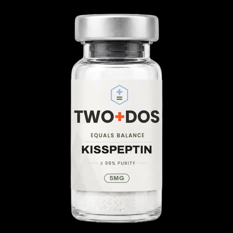
Chemical and physical properties of Kisspeptin-10
| Compound class | RFamide decapeptide; C-terminal fragment of the KISS1 gene product kisspeptin-54 |
|---|---|
| Residue sequence | Tyr-Asn-Trp-Asn-Ser-Phe-Gly-Leu-Arg-Phe-NH2, corresponding to positions 45 through 54 of kisspeptin-54 |
| C-terminal motif | Arg-Phe-NH2 (RFamide); rodent and zebrafish orthologs instead terminate in an RYamide |
| Monoisotopic mass | 1301.6305 Da (PubChem CID 25240297) |
| Computed XLogP | -1.9 (PubChem), consistent with a water-soluble lyophilized solid |
| Topological polar surface area | 538 Ų (PubChem computed) |
| Rotatable bonds | 38 (PubChem computed), reflecting a largely unstructured linear backbone |
| Hydrogen bonding | 18 donors and 16 acceptors (PubChem computed) |
| Cognate receptor | KISS1R, a class A G protein-coupled receptor also catalogued as GPR54, AXOR12 and hOT7T175 |
| Reported binding affinity | Kd 1.9 ± 0.4 nM and Ki 2.33 ± 0.13 nM at human KISS1R; Kd 1.0 ± 0.1 nM at rat KISS1R |
| Principal proteolytic site | The Gly-Leu bond at positions 51 and 52, hydrolyzed by MMP-2, MMP-9, MT1-MMP, MT3-MMP and MT5-MMP |
What is Kisspeptin-10 and where does it sit within the KISS1 precursor?
Kisspeptin-10 occupies the final ten positions of kisspeptin-54 and carries the sequence Tyr-Asn-Trp-Asn-Ser-Phe-Gly-Leu-Arg-Phe-NH2. PubChem records the decapeptide under CID 25240297 with the formula C63H83N17O14, a molecular weight of 1302.4 g/mol and a monoisotopic mass of 1301.6305 Da, catalogued against CAS 374675-21-5.
The KISS1 gene was first characterized as a metastasis suppressor, and the peptide chemistry followed from that. Ohtaki and colleagues reported in Nature in 2001 that KISS1 encodes a 54-residue product they named metastin, identifying it as the ligand of the orphan receptor hOT7T175. Kotani and colleagues independently isolated the same family from human placenta the same year, naming them kisspeptins and matching them to GPR54.
Naming has stayed messy ever since. The identical molecule appears as Kisspeptin-10, KP-10, metastin (45-54) and, because of an alternative numbering convention applied to a shorter precursor fragment, as kisspeptin-13 (4-13). The receptor is variously written KISS1R, GPR54, AXOR12 and hOT7T175. Three shorter processed forms of the precursor, kisspeptin-54, kisspeptin-14 and kisspeptin-13, all terminate in the same ten residues, which is why the decapeptide functions as the common pharmacophore for the whole family.
Two structural features carry the activity. The C-terminal amide is required: the Arg-Phe-NH2 motif places the molecule in the RFamide peptide family, and free-acid analogs lose potency. Species divergence sits at the penultimate position, where rodent and zebrafish orthologs substitute tyrosine for phenylalanine and terminate in an RYamide instead. Chen and colleagues note in a 2025 survey that KISS1R itself is highly conserved across vertebrates, with sequence similarity exceeding 80% in most comparisons.
How does Kisspeptin-10 engage KISS1R at the molecular level?
Kisspeptin-10 binds human KISS1R with a saturation-binding Kd of 1.9 ± 0.4 nM and a competitive Ki of 2.33 ± 0.13 nM, values compiled by Chen and colleagues in 2025. The corresponding rat receptor figures are Kd 1.0 ± 0.1 nM and Ki 1.59 ± 0.07 nM. Calcium flux assays place functional potency at EC50 4.13 ± 0.02 nM for the human receptor.
Downstream coupling runs primarily through Gq/11. Receptor occupancy activates phospholipase C, which hydrolyzes phosphatidylinositol 4,5-bisphosphate into inositol trisphosphate and diacylglycerol, mobilizing intracellular calcium and engaging protein kinase C. Kotani and colleagues reported in 2001 that the isolated kisspeptins triggered calcium mobilization, phosphoinositide turnover, arachidonic acid release and phosphorylation of ERK1/2 and p38, while inhibiting proliferation in the receptor-expressing cell lines used.
Cryo-electron microscopy resolved the interaction in 2024. Shen and colleagues determined the KISS1R complex bound to Kisspeptin-10 at 3.06 Å, deposited as PDB entry 8ZJD, alongside the receptor bound to the synthetic agonist TAK-448 at 3.07 Å as entry 8ZJE. Both peptides adopt a conserved binding pose, and the extracellular loops make the pivotal contacts required for receptor activation.
The same structural work identified a coupling geometry that sets KISS1R apart from other Gq-coupled receptors. Its intracellular transmembrane helix 6 shows an approximately 40° angular deviation relative to comparable receptors, producing a distinct interface with the Gq heterotrimer. Wu and colleagues, publishing in Science Advances in 2024, resolved KISS1R in complex with both Gq and Gi heterotrimers and concluded that the receptor couples to the Gi/o pathway in addition to the better-documented Gq/11 route.
What does the neuroendocrine literature report for Kisspeptin-10?
Kisspeptin-10 acts on KISS1R expressed by gonadotropin-releasing hormone neurons, and the anatomy is unusually tight for a neuropeptide system. Chen and colleagues record that more than 75% of rat GnRH neurons co-express KISS1R messenger RNA, with the peptide itself produced by hypothalamic neurons that also make neurokinin B and dynorphin, the population abbreviated KNDy.
The functional consequence is measurable in human volunteers. George and colleagues reported in 2011 that bolus delivery of Kisspeptin-10 into the circulation of healthy men produced a rapid, amount-dependent rise in serum luteinizing hormone across the range 0.01 to 3.0 µg/kg, with maximal stimulation at 1 µg/kg, taking LH from 4.1 ± 0.4 to 12.4 ± 1.7 IU/liter at 30 minutes. A larger 3 µg/kg bolus produced a smaller response than 1 µg/kg.
Continuous delivery in the same study altered pulse architecture rather than simply raising a mean. Infusion at 4 µg/kg per hour for 22.5 hours raised LH from 5.4 ± 0.7 to 20.8 ± 4.9 IU/liter and testosterone from 16.6 ± 2.4 to 24.0 ± 2.5 nmol/liter. At the lower rate of 1.5 µg/kg per hour, deconvolution analysis resolved an increase in LH pulse frequency from 0.7 ± 0.1 to 1.0 ± 0.2 pulses per hour and secretory burst mass from 3.9 ± 0.4 to 12.8 ± 2.6 IU/liter.
Desensitization is the recurring complication in this literature. Yeung and colleagues addressed it directly in the European Journal of Endocrinology in 2026: continuous delivery across five days left gonadotropins comparable to vehicle even while testosterone stayed elevated, whereas daily eight-hour infusions repeated over twelve days sustained gonadotropin elevation, with the day-12 LH increment smaller than the day-1 increment. Intermittent exposure, in short, held the axis responsive where uninterrupted exposure did not.
Why do Kisspeptin-10 and Kisspeptin-54 behave differently in vivo?
Kisspeptin-10 and kisspeptin-54 share an identical C-terminal decapeptide yet diverge sharply once delivered to an animal, and the difference is largely proteolytic and pharmacokinetic rather than a matter of receptor preference. d'Anglemont de Tassigny and colleagues measured a circulating half-life of roughly 4 minutes for the decapeptide against approximately 32 minutes for the 54-residue form.
The cleavage chemistry was mapped two decades ago. Takino and colleagues reported in Oncogene in 2003 that active MMP-2, MMP-9, MT1-MMP, MT3-MMP and MT5-MMP all hydrolyze the Gly-Leu bond of the KISS1 product, and that the same scissile bond in the isolated decapeptide is cut just as readily. Because that bond sits at positions 51 and 52 of the precursor numbering, it lies inside the shared C-terminal sequence, so every native kisspeptin carries the same liability.
Compartment access separates the two forms as well. In the 2017 comparison, only the 54-residue peptide produced c-Fos activation in GnRH neurons following peripheral delivery, which the authors read as differential access across the blood-brain barrier rather than a difference in intrinsic potency at the receptor. The decapeptide sustained LH release for a far shorter interval than the longer form in the same experiments.
Analog chemistry has been aimed squarely at that Gly-Leu bond. Zhang and colleagues designed phosphinic pseudopeptide analogs intended to resist metalloproteinase hydrolysis and reported in 2018 that one isomer, designated PKPR, retained KISS1R agonist activity measured by ERK1/2 phosphorylation while also inhibiting MMP-2. Robert and colleagues took a different route in 2026, comparing the synthetic analog C6 against both native peptides in HEK293 cells stably expressing the human receptor: C6 produced the most sustained intracellular calcium mobilization and the strongest transcriptional response, but also the most pronounced tachyphylaxis, while kisspeptin-54 alone showed a priming effect on restimulation.
Which non-reproductive systems appear in Kisspeptin-10 studies?
Kisspeptin-10 research extends well beyond the hypothalamic axis, in part because KISS1 entered the literature as a metastasis suppressor rather than a reproductive gene. Ohtaki and colleagues reported that the 54-residue product inhibited chemotaxis and invasion of receptor-transfected CHO cells and attenuated pulmonary metastasis of transfected B16-BL6 melanoma cells in mice.
Oncology work with the decapeptide has continued along that line. Shah and colleagues reported in Scientific Reports in 2025 that exogenous Kisspeptin-10 applied to triple-negative breast cancer cells reduced viability and migration, drove concentration-dependent upregulation of endogenous KISS1 messenger RNA, raised E-cadherin and β-catenin while lowering N-cadherin, CD44 and vimentin, and shifted apoptotic gene expression with BCL2 suppressed. A 2026 report from an overlapping group placed the half-maximal inhibitory concentration at 72.28 nM in ovarian cancer cells and traced activity to an SP1-hTERT-ZEB1 axis with upregulation of miR-200, miR-345 and miR-577.
Skeletal biology produced the most mechanistically specific recent result. Li and colleagues, publishing in Nature Communications in 2024, reported that Kisspeptin-10 binding to Gpr54 in osteoclasts upregulates the phosphatase Dusp18, which dephosphorylates Src at tyrosine 416. Kiss1, Gpr54 and Dusp18 knockout mice each showed osteoclast hyperactivation and bone loss, and the decapeptide suppressed osteoclast activity in vivo.
Vascular and connective-tissue work supplies further mechanism. Radwańska and colleagues reported in Pharmacological Reports in 2026 that Kisspeptin-10 raised glycosaminoglycan content in human cardiac fibroblast cultures in a concentration-dependent manner and increased decorin secretion without altering decorin gene expression, and that the effect required GPR54 and focal adhesion kinase signaling but not phospholipase C, a dissociation from the canonical Gq route. Separate 2025 reports describe barrier and cartilage models: claudin-5 preservation in brain endothelial cells through RhoA/ROCK, claudin-10 upregulation in a rodent stroke model, and attenuation of TNF-α-induced chondrocyte senescence at 50 and 100 nM through SIRT1-dependent suppression of p53 and p21.
What have 2024 to 2026 publications added to the Kisspeptin-10 record?
Kisspeptin-10 publications from 2024 onward cluster into four groups: structural biology of the receptor complex, human-volunteer protocols probing desensitization, analog chemistry aimed at metalloproteinase resistance, and an expanding set of peripheral cell models. The structural entries are the most consequential, because they converted a decade of mutagenesis inference into resolved coordinates.
Radiochemistry entered the record in 2025. Kleynhans and colleagues synthesized a DOTA-conjugated decapeptide intended as a gallium-68 and lutetium-177 chelate, confirmed by receptor stimulation assay that activity was similar for the conjugate and the unmodified peptide, and measured a blood half-life of 18 ± 3 minutes for the radiolabeled construct. That figure is notably longer than the roughly 4 minutes reported for the free peptide, which the conjugation chemistry plausibly explains.
Neuroimmune models are the fastest-growing peripheral application. Kalkan and colleagues reported in Neuropeptides in 2026 that Kisspeptin-10 lowered NLRP3 expression in microglial cells, inhibited caspase-1 and gasdermin D cleavage, reduced IL-1β and IL-18 secretion, and upregulated BAG3, which the authors frame as parallel suppression of the NLRP3-caspase-1-GSDMD axis and activation of BAG3-dependent selective autophagy.
Diagnostic and comparative-physiology reports round out the period. A 2026 study in Clinica Chimica Acta evaluated circulating Kisspeptin-10 as a marker in girls aged 6 to 9 years and found that the composite Kp-10 to basal LH ratio outperformed the individual neuropeptides, with an ROC-derived cut-off below 4.07 ng/mIU giving 72.7% sensitivity, 87.0% specificity and 78% accuracy. Rodent and ovine work over the same window has examined interactions with neurokinin B and dynorphin A at the pituitary level, follicular development through PI3K/AKT/ERK signaling, and pulmonary artery smooth muscle proliferation under hypoxia.
Summary of published research
Findings below are reported as published by the cited authors, in the model systems they used. They describe laboratory research, and none of them characterize use in humans.
Ohtaki T, Shintani Y, Honda S, Matsumoto H, Hori A, Kanehashi K, Terao Y, Kumano S, Takatsu Y, Masuda Y, Ishibashi Y, Watanabe T, Asada M, Yamada T, Suenaga M, Kitada C, Usuki S, Kurokawa T, Onda H, Nishimura O, Fujino M. Metastasis suppressor gene KiSS-1 encodes peptide ligand of a G-protein-coupled receptor. Nature (2001)
- Model system
- cell line (hOT7T175-transfected CHO cells) plus rodent melanoma metastasis model
- Conditions
- purification of the 54-residue KISS1 product from human placenta; chemotaxis and invasion assays in receptor-transfected cells; hOT7T175-transfected B16-BL6 melanoma cells in mice
- Reported finding
- The KISS1 gene product, named metastin, was identified as the endogenous ligand of the orphan receptor hOT7T175; it inhibited chemotaxis and invasion of receptor-transfected CHO cells in vitro and attenuated pulmonary metastasis of transfected B16-BL6 melanoma cells in vivo, establishing the receptor-ligand pairing on which all later work rests.
Kotani M, Detheux M, Vandenbogaerde A, Communi D, Vanderwinden JM, Le Poul E, Brézillon S, Tyldesley R, Suarez-Huerta N, Vandeput F, Blanpain C, Schiffmann SN, Vassart G, Parmentier M. The metastasis suppressor gene KiSS-1 encodes kisspeptins, the natural ligands of the orphan G protein-coupled receptor GPR54. Journal of Biological Chemistry (2001)
- Model system
- cell lines expressing rat and human GPR54
- Conditions
- isolation of natural peptides from human placental extracts; radioligand binding, calcium mobilization, phosphoinositide turnover, arachidonic acid release and kinase phosphorylation assays; tissue expression mapping
- Reported finding
- The isolated kisspeptins bound rat and human GPR54 with low nanomolar affinity and triggered calcium mobilization, phosphoinositide turnover, arachidonic acid release and ERK1/2 and p38 phosphorylation while inhibiting proliferation; receptor transcript was most abundant in placenta, pituitary, pancreas and spinal cord, pointing to endocrine roles beyond the original oncology framing.
Takino T, Koshikawa N, Miyamori H, Tanaka M, Sasaki T, Okada Y, Seiki M, Sato H. Cleavage of metastasis suppressor gene product KiSS-1 protein/metastin by matrix metalloproteinases. Oncogene (2003)
- Model system
- cell-free enzymatic digestion plus cell migration assays
- Conditions
- purified active MMP-2, MMP-9, MT1-MMP, MT3-MMP and MT5-MMP incubated with full-length KISS1 protein and with the isolated decapeptide; migration assays with and without the broad-spectrum inhibitor BB-94
- Reported finding
- All five metalloproteinases cleaved the Gly-Leu peptide bond of both the full-length protein and the decapeptide, and digestion abolished ligand activity; combining the decapeptide with BB-94 produced a synergistic block of cell migration, identifying proteolysis at that single bond as the dominant inactivation route for the whole kisspeptin family.
George JT, Veldhuis JD, Roseweir AK, Newton CL, Faccenda E, Millar RP, Anderson RA. Kisspeptin-10 is a potent stimulator of LH and increases pulse frequency in men. Journal of Clinical Endocrinology and Metabolism (2011)
- Model system
- human volunteers (healthy men)
- Conditions
- bolus delivery into the circulation across 0.01 to 3.0 µg/kg against vehicle; continuous infusion at 4 µg/kg per hour for 22.5 hours and at 1.5 µg/kg per hour, with deconvolution analysis of LH secretion
- Reported finding
- Bolus delivery produced a rapid, amount-dependent LH rise with maximal stimulation at 1 µg/kg (4.1 ± 0.4 to 12.4 ± 1.7 IU/liter at 30 minutes, n = 6), while 3 µg/kg gave a smaller response; infusion at 4 µg/kg per hour raised LH to 20.8 ± 4.9 IU/liter and testosterone from 16.6 ± 2.4 to 24.0 ± 2.5 nmol/liter, and 1.5 µg/kg per hour raised pulse frequency from 0.7 ± 0.1 to 1.0 ± 0.2 pulses per hour with burst mass rising from 3.9 ± 0.4 to 12.8 ± 2.6 IU/liter.
d'Anglemont de Tassigny X, Jayasena CN, Murphy KG, Dhillo WS, Colledge WH. Mechanistic insights into the more potent effect of KP-54 compared to KP-10 in vivo. PLoS One (2017)
- Model system
- rodent (mouse) with parallel circulating half-life measurement
- Conditions
- peripheral delivery of the 10-residue and 54-residue peptides; serial LH sampling, circulating half-life determination and c-Fos immunostaining in GnRH neurons
- Reported finding
- The 54-residue peptide showed a circulating half-life of approximately 32 minutes against roughly 4 minutes for the decapeptide and sustained LH release far longer; only the longer peptide produced c-Fos activation in GnRH neurons after peripheral delivery, which the authors attributed to differential access across the blood-brain barrier rather than a difference in receptor potency.
Robert V, Lomet D, Dardente H, Aucagne V, Beltramo M. The kisspeptin analog C6 elicits greater tachyphylaxis and transcriptional activation than kisspeptin-10 and -54. Molecular and Cellular Endocrinology (2026)
- Model system
- human cell line (HEK293 stably expressing the human kisspeptin receptor)
- Conditions
- three agonists compared head to head, the 10-residue and 54-residue native peptides against the synthetic analog C6; intracellular calcium kinetics, repeat stimulation for tachyphylaxis, and transcriptional profiling
- Reported finding
- C6 produced the most sustained intracellular calcium mobilization and the strongest and most sustained transcriptional response, with upregulation of immediate-early genes, transcription factors and inflammatory mediators, but it also showed the most pronounced tachyphylaxis of the three; the 54-residue peptide alone produced a priming effect on restimulation, separating the three agonists into distinct pharmacological profiles.
Li Z, Yang X, Fu R, Wu Z, Xu S, Jiao J, Qian M, Zhang L, Wu C, Xie T, Yao J, Wu Z, Li W, Ma G, You Y, Chen Y, Zhang HK, Cheng Y, Tang X, Wu P, Lian G, Wei H, Zhao J, Xu J, Ai L, Siwko S, Wang Y, Ding J, Song G, Luo J, Liu M, Xiao J. Kisspeptin-10 binding to Gpr54 in osteoclasts prevents bone loss by activating Dusp18-mediated dephosphorylation of Src. Nature Communications (2024)
- Model system
- rodent (Kiss1, Gpr54 and Dusp18 knockout mice) plus primary osteoclast culture
- Conditions
- genetic deletion of each pathway component with bone phenotyping; Kp-10 delivered in vivo; phosphatase and Src phosphorylation readouts in osteoclasts
- Reported finding
- Kp-10 binding to Gpr54 upregulated the phosphatase Dusp18, which dephosphorylated Src at tyrosine 416 in osteoclasts; Kiss1, Gpr54 and Dusp18 knockout animals each showed osteoclast hyperactivation and bone loss, and Kp-10 suppressed osteoclast activity in vivo, defining a skeletal signaling route distinct from the hypothalamic axis.
Shen S, Wang D, Liu H, He X, Cao Y, Chen J, Li S, Cheng X, Xu HE, Duan J. Structural basis for hormone recognition and distinctive Gq protein coupling by the kisspeptin receptor. Cell Reports (2024)
- Model system
- cryo-electron microscopy of purified receptor-G protein complexes
- Conditions
- KISS1R in complex with Kisspeptin-10 resolved at 3.06 Å (PDB 8ZJD) and with the synthetic agonist TAK-448 at 3.07 Å (PDB 8ZJE), analyzed against other Gq-coupled receptor structures
- Reported finding
- Both peptides adopted a conserved binding pose with the extracellular loops making the contacts pivotal for activation, and the receptor showed an approximately 40° angular deviation in its intracellular transmembrane helix 6 relative to other Gq-coupled receptors, producing a distinct interface with the Gq heterotrimer.
Wu Z, Chen G, Qiu C, Yan X, Xu L, Jiang S, Xu J, Han R, Shi T, Liu Y, Gao W, Wang Q, Li J, Ye F, Pan X, Zhang Z, Ning P, Zhang B, Chen J, Du Y. Structural basis for the ligand recognition and G protein subtype selectivity of kisspeptin receptor. Science Advances (2024)
- Model system
- cryo-electron microscopy of receptor-G protein complexes
- Conditions
- KISS1R-Gq and KISS1R-Gi complexes bound to the synthetic agonist TAK-448, plus a KISS1R-Gq complex bound to kisspeptin-54, compared for interface geometry
- Reported finding
- The structures showed that KISS1R couples to the Gi/o pathway in addition to the established Gq/11 route, and identified conformational differences in the receptor loops that account for G protein subtype selectivity, extending the coupling picture beyond the single-pathway model previously assumed for this receptor.
Kleynhans J, Reeve R, Driver CHS, et al. Synthesis and characterisation of DOTA-kisspeptin-10 as a potential gallium-68/lutetium-177 pan-tumour radiopharmaceutical. Journal of Neuroendocrinology (2025)
- Model system
- chemical synthesis with receptor stimulation assay and rodent blood clearance
- Conditions
- DOTA chelator conjugated to the decapeptide; receptor activation compared against the unmodified peptide; radiolabeled construct tracked for blood clearance
- Reported finding
- Receptor stimulation was similar for the DOTA conjugate and the unmodified decapeptide, indicating that chelator conjugation preserved receptor engagement, and the radiolabeled construct cleared rapidly from blood with a measured half-life of 18 ± 3 minutes, establishing a starting point for gallium-68 and lutetium-177 analog development.
Shah H, Mohan AM, Buch L, Ramachandran AV, Pandya P. Exogenous kisspeptin-10 treatment shows pleiotropy via induction of KISS1 expression, metastasis suppression, and promotes apoptosis in triple-negative breast cancer. Scientific Reports (2025)
- Model system
- human cell line (triple-negative breast cancer)
- Conditions
- exogenous decapeptide applied across a concentration range; viability and migration assays, KISS1 transcript quantification, epithelial and mesenchymal marker panels, apoptotic gene expression
- Reported finding
- The peptide reduced viability and migration and produced concentration-dependent upregulation of endogenous KISS1 messenger RNA; E-cadherin and β-catenin rose while N-cadherin, CD44 and vimentin fell, reversing the mesenchymal marker profile, and pro-apoptotic gene expression increased with BCL2 suppressed.
Yeung AC, Phylactou M, Koysombat K, Tsoutsouki J, Nyunt SW, Young M, Patel AH, Daniels E, Pierret ACS, Mills EG, Comninos AN, Abbara A, Dhillo WS. Chronic subcutaneous kisspeptin-10 stimulates gonadotropin secretion for 12 days in healthy men. European Journal of Endocrinology (2026)
- Model system
- human volunteers (healthy men)
- Conditions
- three protocols compared: acute amount-response infusions, continuous infusion across five days, and daily eight-hour infusions repeated over twelve days, each against vehicle
- Reported finding
- Acute infusions produced amount-dependent increases in LH, FSH and testosterone; continuous five-day delivery left gonadotropins comparable to vehicle despite maintained testosterone elevation, whereas the intermittent twelve-day protocol sustained gonadotropin elevation with a smaller LH increment on day 12 than on day 1, indicating that exposure pattern rather than total exposure governs whether the axis stays responsive.
What laboratory handling information is published?
Kisspeptin-10 is supplied as a lyophilized powder, and its computed profile matches that format. PubChem lists an XLogP of -1.9, a topological polar surface area of 538 Ų, eighteen hydrogen bond donors and sixteen acceptors for CID 25240297, describing a strongly polar linear peptide with 38 rotatable bonds and no meaningful lipophilic character. Aqueous buffer and culture medium are the vehicles used across the published methods.
Metalloproteinase susceptibility is the governing practical constraint. Because MMP-2, MMP-9, MT1-MMP, MT3-MMP and MT5-MMP all hydrolyze the Gly-Leu bond at positions 51 and 52, any matrix carrying active metalloproteinase activity, including serum-supplemented medium and conditioned medium from invasive cell lines, will degrade the peptide over the course of an experiment. Published protocols that need extended exposure either replenish the peptide, co-apply a broad-spectrum metalloproteinase inhibitor as Takino and colleagues did with BB-94, or switch to a protease-resistant analog such as the phosphinic pseudopeptide PKPR.
Working concentrations in the cited literature span a narrow band relative to many research peptides, because the receptor affinity is high. Binding sits at low nanomolar (Kd 1.9 ± 0.4 nM at the human receptor) and calcium-flux potency at EC50 4.13 ± 0.02 nM, and recent cell studies reflect that: 50 and 100 nM in chondrocyte senescence work, a half-maximal inhibitory concentration of 72.28 nM in ovarian cancer cells. Rodent protocols are quoted per kilogram, for example 50 nmol/kg in a blood-brain barrier model, and human-volunteer protocols in micrograms per kilogram per hour.
Identity confirmation follows standard peptide practice. Electrospray mass spectrometry is run against a monoisotopic mass of 1301.6305 Da, giving a singly protonated ion near m/z 1302.6 and a doubly protonated ion near m/z 651.8, with the doubly charged species usually dominating at this mass. Purity is determined by reversed-phase HPLC and reported on the lot certificate of analysis. A radioiodinated form, [125I]kisspeptin-10, is catalogued among the PubChem synonyms and has served as the tracer in saturation and competition binding studies at both rat and human receptors.
Lyophilized material is normally held frozen, protected from light and moisture, with aqueous stock solutions aliquoted so repeated freeze-thaw cycles are avoided. The C-terminal amide is load-bearing for activity, so conditions favoring deamidation or C-terminal hydrolysis are the storage failure mode that matters most for this molecule. This material is offered for laboratory research use only. It is not a drug, not a food, and is not intended for human or veterinary use.
Frequently asked research questions
What is the difference between Kisspeptin-10 and kisspeptin-54?
Kisspeptin-54 is the 54-residue product of the human KISS1 gene, and Kisspeptin-10 is its final ten residues, positions 45 through 54. Both engage KISS1R through the same C-terminal sequence, but the reported circulating half-life differs sharply, roughly 4 minutes for the decapeptide against approximately 32 minutes for the longer form.
Why is the C-terminal amide important in Kisspeptin-10?
The molecule terminates in Arg-Phe-NH2, which places it in the RFamide peptide family and is required for potency at KISS1R. Rodent and zebrafish orthologs substitute tyrosine at the penultimate position and terminate in an RYamide instead, so the human decapeptide and its rodent counterpart are chemically distinct entities despite sharing the same numbering.
Which receptor does Kisspeptin-10 bind, and how tightly?
The cognate receptor is KISS1R, catalogued in older papers as GPR54, AXOR12 and hOT7T175. Compiled values put the saturation-binding Kd at 1.9 ± 0.4 nM and the competitive Ki at 2.33 ± 0.13 nM for the human receptor, with calcium-flux potency at EC50 4.13 ± 0.02 nM. Rat receptor affinity is roughly twofold higher.
What does the Gly-Leu cleavage site mean for experimental design?
Matrix metalloproteinases hydrolyze the Gly-Leu bond at positions 51 and 52, and that bond lies inside the shared C-terminal sequence of every native kisspeptin. Digestion abolishes ligand activity, so experiments run in metalloproteinase-rich matrices need peptide replenishment, an inhibitor co-application, or a protease-resistant analog.
Which model systems dominate the Kisspeptin-10 literature?
Four recur across the citations above: human-volunteer neuroendocrine protocols with serial hormone sampling, rodent knockout and challenge models, human and murine cell lines spanning cancer, endothelium, chondrocytes, cardiac fibroblasts and microglia, and purified receptor preparations for binding assays and cryo-electron microscopy. Structural coordinates for the receptor complex became available in 2024.
Kisspeptin-10 at TWO+DOS
TWO+DOS supplies Kisspeptin-10 as a research-use-only compound, third-party tested, with certificates of analysis emailed immediately on request.
View the Kisspeptin (5mg)listing →Related research overviews
References
- PubChem CID 25240297: Kisspeptin-10 compound summary
- Metastasis suppressor gene KiSS-1 encodes peptide ligand of a G-protein-coupled receptor (Nature 2001)
- The metastasis suppressor gene KiSS-1 encodes kisspeptins, the natural ligands of the orphan G protein-coupled receptor GPR54 (J Biol Chem 2001)
- Cleavage of metastasis suppressor gene product KiSS-1 protein/metastin by matrix metalloproteinases (Oncogene 2003)
- Kisspeptin-10 is a potent stimulator of LH and increases pulse frequency in men (J Clin Endocrinol Metab 2011)
- Mechanistic insights into the more potent effect of KP-54 compared to KP-10 in vivo (PLoS One 2017)
- Functional examination of novel kisspeptin phosphinic peptides (PLoS One 2018)
- Kisspeptin-10 binding to Gpr54 in osteoclasts prevents bone loss by activating Dusp18-mediated dephosphorylation of Src (Nat Commun 2024)
- Structural basis for hormone recognition and distinctive Gq protein coupling by the kisspeptin receptor (Cell Rep 2024)
- Structural basis for the ligand recognition and G protein subtype selectivity of kisspeptin receptor (Sci Adv 2024)
- Kisspeptin Receptor Agonists and Antagonists: Strategies for Discovery and Implications for Human Health and Disease (Int J Mol Sci 2025)
- Chronic subcutaneous kisspeptin-10 stimulates gonadotropin secretion for 12 days in healthy men (Eur J Endocrinol 2026)
For research use only. Not for human or veterinary use. Not for diagnostic or therapeutic use.