
MOTS-c: Mitochondrial Microprotein Structure and Signalling Mechanism
By the TWO+DOS Research Team · Published 2026-08-12
For research use only. Not for human or veterinary use. Not for diagnostic or therapeutic use.
MOTS-c, the mitochondrial open reading frame of the 12S rRNA type-c, is a 16-residue microprotein of 2174.6 g/mol encoded by a 51-base-pair reading frame inside mitochondrial DNA. Its sequence, MRWQEMGYIFYPRKLR, places it in the mitochondrial-derived peptide family alongside humanin, and it was first described in 2015.
What makes the molecule unusual is its address rather than its size. Mitochondrial DNA had been understood to encode components of the respiratory chain and the RNAs needed to build them. MOTS-c is a mitochondrially encoded factor that acts on the nuclear genome, reversing the assumed direction of organelle-to-nucleus instruction. This overview covers the reading frame, the folate-cycle route to AMPK, the nuclear work, the sequence variant, and how the peptide is measured.
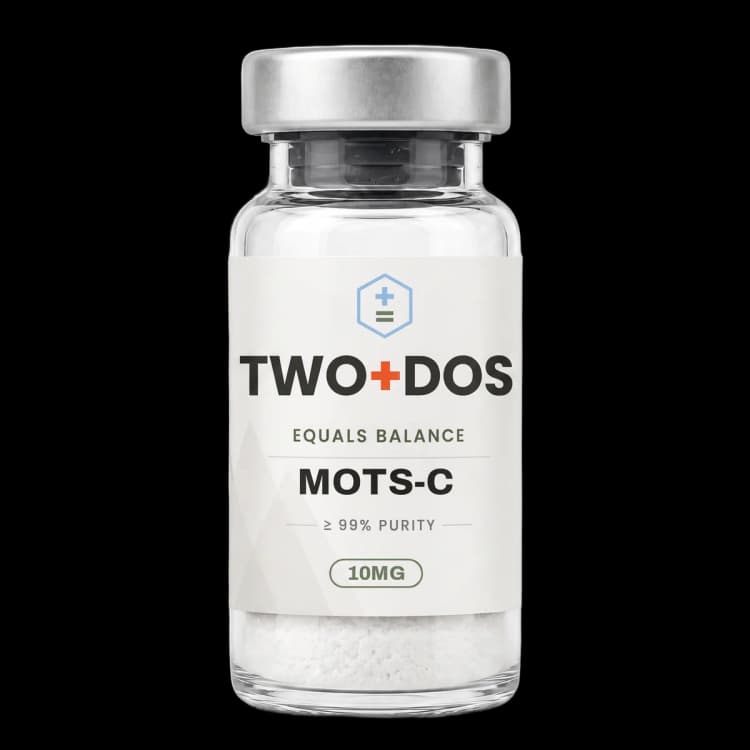
Chemical and physical properties of MOTS-c
| Peptide class | 16-residue mitochondrial-derived microprotein |
|---|---|
| Amino acid sequence | MRWQEMGYIFYPRKLR, listed in PubChem as H-MRWQEMGYIFYPRKLR-OH |
| Molecular formula | C101H152N28O22S2 |
| Molecular mass | 2174.6 g/mol; exact mass 2174.111 Da; monoisotopic mass 2173.108 Da |
| CAS number | 1627580-64-6 |
| PubChem CID | 146675088 |
| Other registry identifiers | UNII A5CV6JFB78; UniProtKB A0A0C5B5G6, entry name MOTSC_HUMAN, gene MT-RNR1 |
| InChIKey | WYTHCOXVWRKRAH-LOKRTKBUSA-N |
| Genomic origin | 51-base-pair short open reading frame within the mitochondrial 12S rRNA region, mtDNA positions m.1343 to m.1393 |
| Sulfur content | Two sulfur atoms, both from methionine at positions 1 and 6. No cysteine, so no disulfide bridge is possible |
| Computed polarity descriptors | XLogP minus 3.9; topological polar surface area 890 square angstroms; 31 hydrogen-bond donors; 29 acceptors; 73 rotatable bonds (PubChem computed) |
What is MOTS-c and where is it encoded?
MOTS-c is a 16-residue microprotein translated from a 51-base-pair short open reading frame located inside the mitochondrial 12S ribosomal RNA gene, MT-RNR1, spanning mitochondrial DNA positions m.1343 to m.1393. Lee and colleagues reported the sequence in Cell Metabolism in 2015, naming it for the reading frame it occupies.
One production detail is often blurred. Although the reading frame sits in mitochondrial DNA, the peptide is understood to be translated in the cytoplasm using the universal genetic code, because tandem start and stop codons under the mitochondrial code would not yield the observed 16-residue product. The gene is mitochondrial; the ribosome is not. Review work describes the sequence as conserved across 14 species, the first 11 residues most strongly.
The physical chemistry follows from the sequence. The formula C101H152N28O22S2 carries two sulfur atoms, both from the methionine residues at positions 1 and 6, and no cysteine appears in the chain, so disulfide chemistry is not a consideration. PubChem computes an XLogP of minus 3.9 and a topological polar surface area of 890 square angstroms, describing a small, strongly polar peptide that does not cross lipid bilayers readily.
How does MOTS-c connect to AMPK?
MOTS-c reaches AMP-activated protein kinase indirectly. Lee and colleagues reported in 2015 that the peptide inhibits the folate cycle and the de novo purine biosynthesis pathway tethered to it. Blocked purine synthesis raises intracellular AICAR, an AMP mimetic that binds and activates AMPK, with skeletal muscle identified as the principal target tissue.
The metabolomic signature reported in that work is specific and testable. Cells exposed to the peptide showed a concerted fall in 5-methyl-tetrahydrofolate, the most abundant activated folate, a fall in methionine, and a rise in homocysteine. Expression changes across folate-methionine cycle and purine synthesis enzymes were detectable four hours after exposure.
Downstream of AMPK the literature converges on the PGC-1alpha axis. Gudiksen and colleagues tested the dependency in Free Radical Biology and Medicine in 2026 using two transgenic mouse strains, and reported that augmented skeletal muscle mitochondrial bioenergetic capacity required both PGC-1alpha and AMPK. Reactive oxygen species emission and oxidation-related protein damage fell with no change in mitochondrial respiratory protein content, pointing to a change in the intrinsic quality of existing mitochondria rather than in mitochondrial volume.
Cardiac tissue supplies a numerical example. In high-resolution respirometry published by Pham and colleagues in 2025, diabetic rat mitochondria from animals not receiving the peptide showed oxygen flux in the complex I plus complex II oxidative phosphorylation state 13 percent below control, a difference absent in the exposed group. That same group showed elevated hydrogen peroxide production at the leak state, running counter to the redox findings in skeletal muscle.
What happens when MOTS-c enters the nucleus?
MOTS-c translocates from the cytoplasm into the nucleus under metabolic stress. Kim and colleagues showed in Cell Metabolism in 2018 that the movement is AMPK-dependent, and that nuclear MOTS-c regulates a broad set of genes during glucose restriction, including genes carrying antioxidant response elements, while interacting with stress-responsive transcription factors such as NFE2L2/NRF2.
The significance of that paper sits in its framing. Nuclear factors had long been known to govern the mitochondrial genome; the 2018 work established traffic in the opposite direction, with a mitochondrially encoded peptide binding promoter regions of nuclear genes. Later review work adds activating transcription factors 1 and 7.
The NRF2 connection recurs across recent mechanistic work. Zhang and colleagues reported in Redox Biology in 2026 that an engineered analogue acted through increased nuclear NRF2 translocation, with the effect abolished under NRF2 inhibition and in NRF2-knockout animals. Li and colleagues reported in Life Sciences in 2026 that the peptide appears to interact with KEAP1, the sensor that holds NRF2 inactive, maintaining the KEAP1-PGAM5 interaction and blocking downstream AIFM1 nuclear translocation; KEAP1 overexpression abolished the effect.
Does MOTS-c act only through AMPK?
MOTS-c has documented actions that do not require AMPK. Jia and colleagues reported in Theranostics in 2024 that the peptide binds the carboxy-terminus of TRIM72, a plasma-membrane repair protein, and accelerates its trafficking to damaged membrane. Knockdown of TRIM72 by siRNA blocked the effect, while an AMPK inhibitor did not.
That paper also identified a direct lipid interaction. Dot blot analysis showed binding to phosphatidylinositol 4,5-bisphosphate, and in TRIM72-knockout mice the peptide still increased fusion of vesicles with the membrane, indicating a second route independent of the protein partner. On its own it had negligible effect on membrane repair, making it a facilitator of TRIM72 traffic rather than a repair agent itself.
Cell entry remains open. Published work describes intracellular targets rather than a characterized cell-surface receptor, and the high polarity of the molecule is described as a limiting factor for membrane permeability. Zhang and colleagues addressed this in 2026 by substituting alanine for the arginine at position 13, raising computed hydrophobicity from minus 0.938 to minus 0.544, and showed that the R13A analogue enters cells through LAT1-mediated transport.
Context also changes the result. Xing and colleagues reported in Inflammation and Regeneration in 2026 that human mesenchymal stromal cells from donors at or above 30 kg per square metre body mass index expressed less of the peptide than cells from donors below that threshold, and that supplying it activated AMPK signalling while reducing proliferation and raising p16, p21 and TNF-alpha expression. Metabolic signalling and reparative function came apart in that system.
What is the m.1382A>C K14Q variant of MOTS-c?
The m.1382A>C polymorphism in mitochondrial DNA replaces the lysine at position 14 of MOTS-c with glutamine, producing the variant designated K14Q. Fuku and colleagues described the variation in Aging Cell in 2015 as specific to Northeast Asian populations and carried on the ancestral haplogroup D4b2, which had been reported in association with exceptional lifespan in Japanese cohorts.
Zempo and colleagues tested the functional consequences in Aging in 2021. Meta-analysis of the J-MICC, MEC and TMM cohorts, totalling 27,527 individuals, associated the C allele with higher prevalence of type 2 diabetes in males but not females, and described the substitution as altering peptide structure in a way that reduces insulin-sensitizing activity. An expanded analysis in 736 individuals showed no effect on lifespan. For laboratory work the variant is a naturally occurring single-residue structure-activity probe, and a genotype that varies by ancestry, which can confound human sample sets.
How is MOTS-c quantified in biological samples?
MOTS-c is quantified in plasma either by immunoassay or by liquid chromatography coupled to mass spectrometry. Knoop, Thomas and Thevis validated an LC/MS method against World Anti-Doping Agency laboratory standards in Rapid Communications in Mass Spectrometry in 2019, reporting a lower limit of detection of 100 pg/mL for the human peptide, alongside four metabolites and two oxidation products.
That paper contains a discrepancy anyone reading this literature should know about. Endogenous reference concentrations obtained by ELISA sat between 45.9 and 218.5 ng/mL, while the mass spectrometric method did not return values consistent with that range. The authors flagged the mismatch as warranting further investigation. Immunoassay-derived concentrations are method-specific numbers, and results from different platforms should not be pooled.
Relative change is measured more consistently than absolute level. Reynolds and colleagues reported in Nature Communications in 2021 that in 10 sedentary males performing stationary cycling, skeletal muscle expression rose 11.9-fold immediately after the session, while the circulating pool rose 1.6-fold during and 1.5-fold after, returning to baseline within four hours. Gudiksen and colleagues added a sourcing observation in 2026: interstitial concentrations rose during exercise, but no arterio-venous difference across the limb was detectable.
Summary of published research
Findings below are reported as published by the cited authors, in the model systems they used. They describe laboratory research, and none of them characterize use in humans.
Lee C, Zeng J, Drew BG, Sallam T, Martin-Montalvo A, et al. The mitochondrial-derived peptide MOTS-c promotes metabolic homeostasis and reduces obesity and insulin resistance. Cell Metabolism (2015)
- Model system
- Cell line and rodent
- Conditions
- Metabolomic and transcriptional profiling of exposed cells; mouse studies under age-associated and high-fat-diet conditions
- Reported finding
- A 16-amino-acid peptide was identified and named. Cellular actions inhibited the folate cycle and the tethered de novo purine biosynthesis pathway, leading to AMPK activation, with skeletal muscle the apparent primary target organ. In mice, insulin resistance associated with age and with a high-fat diet was reduced.
Kim KH, Son JM, Benayoun BA, Lee C. The Mitochondrial-Encoded Peptide MOTS-c Translocates to the Nucleus to Regulate Nuclear Gene Expression in Response to Metabolic Stress. Cell Metabolism (2018)
- Model system
- Cell line
- Conditions
- Glucose restriction as the stressor with AMPK inhibition to test dependency; promoter occupancy and transcription-factor interaction assays
- Reported finding
- The peptide translocated to the nucleus and regulated nuclear gene expression in an AMPK-dependent manner, acting on genes including those carrying antioxidant response elements, and interacting with stress-responsive transcription factors such as NFE2L2/NRF2.
Knoop A, Thomas A, Thevis M. Development of a mass spectrometry based detection method for the mitochondrion-derived peptide MOTS-c in plasma samples for doping control purposes. Rapid Communications in Mass Spectrometry (2019)
- Model system
- Analytical method development in human plasma
- Conditions
- LC/MS validated to the World Anti-Doping Agency International Standard for Laboratories; long-term stability and in vitro metabolism evaluated
- Reported finding
- A lower limit of detection of 100 pg/mL was achieved in plasma, with four metabolites and two oxidation products implemented. Endogenous reference concentrations measured by ELISA, 45.9 to 218.5 ng/mL, diverged substantially from the mass spectrometric results, a discrepancy the authors flagged for further investigation.
Reynolds JC, Lai RW, Woodhead JST, Joly JH, Mitchell CJ, et al. MOTS-c is an exercise-induced mitochondrial-encoded regulator of age-dependent physical decline and muscle homeostasis. Nature Communications (2021)
- Model system
- Rodent and human
- Conditions
- Mice at 2, 12 and 22 months; human arm of 10 sedentary males performing stationary cycling, with muscle biopsy and circulating sampling at four timepoints
- Reported finding
- In humans, skeletal muscle expression rose 11.9-fold immediately after the session while the circulating pool rose 1.6-fold during and 1.5-fold after, returning to baseline by four hours. In mice the peptide regulated nuclear genes related to metabolism and proteostasis, and myoblast adaptation to metabolic stress.
Zempo H, Kim SJ, Fuku N, Nishida Y, Higaki Y, et al. A pro-diabetogenic mtDNA polymorphism in the mitochondrial-derived peptide, MOTS-c. Aging (Albany NY) (2021)
- Model system
- Human population genetics, meta-analysis
- Conditions
- Meta-analysis across the J-MICC, MEC and TMM cohorts totalling 27,527 individuals; separate lifespan analysis in 736 individuals
- Reported finding
- Males carrying the C allele of m.1382A>C, which yields the K14Q substitution, showed higher prevalence of type 2 diabetes; females did not. The substitution was described as altering peptide structure in a way that reduces insulin-sensitizing activity. The expanded analysis showed no effect on lifespan.
Jia H, Zhou LC, Chen YF, Zhang W, Qi W, et al. Mitochondria-encoded peptide MOTS-c participates in plasma membrane repair by facilitating the translocation of TRIM72 to membrane. Theranostics (2024)
- Model system
- Cell line and rodent
- Conditions
- Hypotonic membrane damage in vitro with siRNA-TRIM72 and AMPK inhibitor controls; co-immunoprecipitation; dot blot lipid binding; TRIM72-knockout mice
- Reported finding
- The peptide interacted with the TRIM72 carboxy-terminus but not the amino-terminus and accelerated TRIM72 trafficking to injured membrane. The effect was blocked by siRNA-TRIM72 but not by an AMPK inhibitor. Direct binding to phosphatidylinositol 4,5-bisphosphate was detected, and vesicle fusion still increased in TRIM72-knockout mice.
Pham T, Taberner A, Hickey A, Han JC. Mitochondria-derived peptide MOTS-c restores mitochondrial respiration in type 2 diabetic heart. Frontiers in Physiology (2025)
- Model system
- Rodent, isolated cardiac mitochondria
- Conditions
- Male Wistar rats, 15-week high-fat diet at 43 percent digestible lipid with low-level streptozotocin at week 8; peptide at 15 mg/kg daily by intraperitoneal route for 3 weeks; respirometry with Magnesium Green and Amplex UltraRed
- Reported finding
- Oxygen flux in the complex I plus complex II oxidative phosphorylation state was 13 percent lower in diabetic mitochondria from animals not receiving the peptide, and this difference was absent in the exposed group. Citrate synthase activity rose and ATP hydrolysis during anoxia fell in the exposed group, while hydrogen peroxide production at the leak state was elevated.
Gudiksen A, Hansen CC, van der Stede T, Daugaard AH, Schmidt JH, et al. MOTS-c improves intrinsic muscle mitochondrial bioenergetic health and efficiency in a PGC-1alpha/AMPK-dependent manner. Free Radical Biology and Medicine (2026)
- Model system
- Rodent (two transgenic strains) and human
- Conditions
- PGC-1alpha and AMPK transgenic mouse models with respirometry, reactive oxygen species emission, protein-damage markers and RNA sequencing; human interstitial and arterio-venous sampling during exercise
- Reported finding
- Augmented muscle mitochondrial bioenergetic capacity required both PGC-1alpha and AMPK. Reactive oxygen species emission and oxidation-related protein damage were lower with no change in mitochondrial respiratory protein content, indicating intrinsic rather than volumetric change. In humans, interstitial concentrations rose with no arterio-venous difference detected.
Xing L, Lu B, Zhu X, Al Saeedi M, Lerman A, Eirin A, Cohen P, Lerman LO. Mitochondrial-derived peptide MOTS-c activates metabolic signaling but blunts reparative function in human mesenchymal stromal cells. Inflammation and Regeneration (2026)
- Model system
- Human primary cells and rodent
- Conditions
- Mesenchymal stromal cells from donors at or above 30 kg per square metre body mass index versus donors below it; exogenous peptide exposure; in vivo kidney injury model
- Reported finding
- Basal expression was lower in cells from the higher body mass index group. Exogenous peptide restored intracellular levels and activated AMPK signalling, yet reduced proliferation and raised p16, p21 and TNF-alpha expression. Cells exposed beforehand did not restore renal perfusion, fibrosis or tubular measures, and the same exposure reduced the reparative efficacy of cells from the lower-index group.
Zhang YL, Huang G, Li SP, Zhang WL, Chen D, et al. LAT1-mediated delivery of engineered R13A-MOTS-c attenuates radiation-induced lung injury via Nrf2 activation and mitochondrial protection. Redox Biology (2026)
- Model system
- Cell line and rodent
- Conditions
- Arginine-to-alanine substitution at position 13; uptake assays with LAT1 inhibition; mice receiving 20 Gy thoracic irradiation with the analogue at 5 mg/kg daily for 2 weeks; NRF2-knockout controls
- Reported finding
- The R13A substitution raised computed hydrophobicity from minus 0.938 to minus 0.544 and increased cellular uptake, which proceeded through LAT1-mediated transport. The analogue reduced inflammatory signalling, oxidative damage and mitochondrial impairment in lung cells and in irradiated mice via increased nuclear NRF2 translocation. Effects were lost under LAT1 inhibition, under NRF2 inhibition and in NRF2-knockout animals.
What laboratory handling information is published?
MOTS-c is supplied as a lyophilized solid. Peer-reviewed handling data is thin, and most storage and solubility figures in circulation come from supplier specifications rather than published measurement, so they belong in a method section labelled as such. The peer-reviewed material that exists concerns analytical stability: Knoop and colleagues evaluated long-term stability and in vitro metabolism while validating their assay.
The chemistry points to one specific liability. Methionine occupies positions 1 and 6 and no cysteine appears in the chain, so methionine sulfoxide formation is the oxidation route to anticipate and disulfide scrambling is not. Purity and stability assessments should resolve the oxidized species explicitly rather than reporting a single main-peak percentage.
Solution behaviour follows from the computed descriptors. An XLogP of minus 3.9 and a topological polar surface area of 890 square angstroms describe a strongly hydrophilic molecule, and the 2026 Redox Biology work states that membrane permeability is poor because of that polarity, which is why an alanine substitution at position 13 was engineered. Groups studying uptake should expect the unmodified sequence to enter cells inefficiently and should include an uptake control.
Frequently asked research questions
What does the name MOTS-c stand for, and is the peptide made by mitochondrial ribosomes?
Mitochondrial open reading frame of the 12S rRNA type-c, named for a 51-base-pair reading frame inside the gene MT-RNR1 at mtDNA positions m.1343 to m.1393. Translation is understood to occur in the cytoplasm using the universal genetic code, because tandem start and stop codons under the mitochondrial code would not give the observed 16-residue peptide.
Does MOTS-c have a known cell-surface receptor?
No characterized cell-surface receptor appears in the mechanistic work reviewed here. Documented targets are intracellular: the folate-methionine cycle and de novo purine synthesis upstream of AMPK, NFE2L2/NRF2, KEAP1, TRIM72, and the membrane lipid phosphatidylinositol 4,5-bisphosphate. Entry of an engineered R13A analogue has been attributed to LAT1-mediated transport.
Why do reported circulating concentrations vary so widely between studies?
Platform. Knoop and colleagues reported an ELISA-derived endogenous range of 45.9 to 218.5 ng/mL alongside a validated LC/MS method with a 100 pg/mL detection limit, and the two did not agree. Immunoassay values are method-specific and should not be pooled across platforms.
What is the K14Q variant of MOTS-c?
A naturally occurring substitution produced by the mtDNA polymorphism m.1382A>C, replacing lysine 14 with glutamine. It is specific to Northeast Asian populations on haplogroup D4b2. A meta-analysis of 27,527 individuals associated the C allele with higher type 2 diabetes prevalence in males and described the variant as structurally altered with reduced insulin-sensitizing activity.
MOTS-c at TWO+DOS
TWO+DOS supplies MOTS-c as a research-use-only compound, third-party tested, with certificates of analysis emailed immediately on request.
View the MOTS-Clisting →Related research overviews
References
- PubChem CID 146675088: formula, mass and computed descriptors
- UniProtKB A0A0C5B5G6 (MOTSC_HUMAN): sequence and gene
- Lee et al. 2015, Cell Metabolism (PMID 25738459)
- Kim et al. 2018, Cell Metabolism (PMID 29983246)
- Knoop et al. 2019, Rapid Commun Mass Spectrom (PMID 30394592)
- Reynolds et al. 2021, Nature Communications (PMID 33473109)
- Zempo et al. 2021, Aging (PMID 33468709)
- Fuku et al. 2015, Aging Cell: mtDNA coordinates
- Jia et al. 2024, Theranostics (PMID 39267782)
- Pham et al. 2025, Frontiers in Physiology (PMID 40661667)
- Gudiksen et al. 2026, Free Radic Biol Med (PMID 41520850)
- Zhang et al. 2026, Redox Biology (PMID 42142418)
For research use only. Not for human or veterinary use. Not for diagnostic or therapeutic use.