
SS-31 (Elamipretide): Cardiolipin Binding and Mitochondrial Membrane Research
By the TWO+DOS Research Team · Published 2026-08-12
For research use only. Not for human or veterinary use. Not for diagnostic or therapeutic use.
SS-31, known in the published literature as elamipretide, is a synthetic aromatic-cationic tetrapeptide with the sequence D-Arg-Dmt-Lys-Phe-NH2. Research characterizes it as a molecule that associates with cardiolipin, the signature phospholipid of the inner mitochondrial membrane, and mechanistic work centers on membrane electrostatics rather than bulk radical scavenging.
This overview collects what peer-reviewed sources report for the molecule: verified chemical identifiers, the biophysical measurements behind its cardiolipin selectivity, the mitochondrial proteins it has been cross-linked to, and results from cell-free, cell-line and rodent model systems published between 2004 and 2026. All material described here is for laboratory research use only.
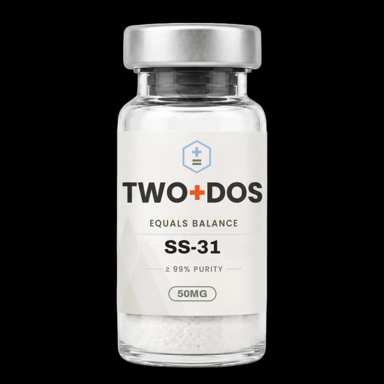
Chemical and physical properties of SS-31 (Elamipretide)
| Compound class | Aromatic-cationic tetrapeptide (Szeto-Schiller series) |
|---|---|
| Residue sequence | D-Arg-Dmt-Lys-Phe-NH2, where Dmt is 2',6'-dimethyltyrosine |
| Non-standard residues | D-configured arginine at position 1; dimethylated tyrosine at position 2 |
| C-terminus | Primary carboxamide (Phe-NH2), not a free acid |
| Formal charge | +3 at neutral pH (Mitchell et al., J Biol Chem 2020) |
| Monoisotopic mass | 639.3857 Da (PubChem CID 11764719) |
| Computed XLogP | 0 (PubChem), consistent with a highly polar molecule |
| Topological polar surface area | 267 Ų (PubChem computed) |
| Rotatable bonds | 19 (PubChem computed) |
| Reported membrane target | Cardiolipin of the inner mitochondrial membrane |
| Reported bilayer affinity | KD 2.0-2.9 µM for 20 mol% tetraoleoyl-cardiolipin vesicles |
What structural features define the SS-31 tetrapeptide?
SS-31 is a four-residue peptide in which cationic and aromatic side chains alternate: D-arginine, 2',6'-dimethyltyrosine, lysine and phenylalanine, capped as a C-terminal primary amide. PubChem records the molecular formula as C32H49N9O5 with a molecular weight of 639.8 g/mol and a monoisotopic mass of 639.3857 Da under CID 11764719.
Two design choices separate SS-31 from a conventional all-L peptide. The arginine at position 1 is in the D configuration, a substitution the Szeto-Schiller series was built around because non-natural stereochemistry at the N-terminus resists aminopeptidase cleavage. Position 2 carries 2',6'-dimethyltyrosine rather than tyrosine, adding steric bulk and aromatic character to the phenol ring.
Charge distribution follows from that architecture. The free N-terminal amine, the guanidinium of D-arginine and the lysine side chain each carry a positive charge at neutral pH, while the C-terminal amide removes the anionic carboxylate that a free acid would contribute. Mitchell and colleagues describe the resulting molecule as carrying a formal charge of +3 at neutral pH. PubChem computes an XLogP of 0 and a topological polar surface area of 267 Ų, values consistent with a strongly polar, non-lipophilic compound that nevertheless partitions onto membranes.
How does SS-31 associate with cardiolipin in the inner mitochondrial membrane?
SS-31 binds anionic lipid bilayers, with a marked preference for cardiolipin. In model membranes containing 20 mol% tetraoleoyl-cardiolipin, isothermal titration calorimetry returned dissociation constants of 2.0-2.9 µM, compared with 6.0-13 µM for bilayers containing the monoanionic lipid phosphatidylglycerol at the same mole fraction.
Partition coefficients from the same 2020 study separate the two lipid classes further: roughly 5.07 × 10⁴ for cardiolipin-containing vesicles and 5.90 × 10⁴ for monolyso-cardiolipin, against 1.28 × 10⁴ for phosphatidylglycerol. Enthalpy changes were small (about −4.2 kJ mol⁻¹ for cardiolipin), while the entropic term dominated at roughly −22.7 kJ mol⁻¹, indicating an association driven mainly by entropy rather than by strong enthalpic contacts.
The consequence measured at the bilayer surface is electrostatic. Zeta potential measurements showed a saturable reduction in the magnitude of surface charge as peptide was added, plateauing near −30 mV, so the membrane remained net negative even at binding saturation. Molecular dynamics and NMR placed the basic side chains at the interfacial region tethered to headgroup phosphates, with aromatic rings only partially buried, and small-angle X-ray scattering detected no major topological alteration of the bilayer even at the highest peptide-to-lipid ratios tested.
Cardiolipin makes this selectivity biologically specific. The same source notes that cardiolipin comprises roughly 10-20 mol% of total lipid in the inner mitochondrial membrane and is largely confined to that compartment. Exogenously added SS peptides were reported to cross the plasma membrane by a nonsaturable route that does not require metabolic input, then to accumulate 1000-5000 fold at the inner mitochondrial membrane.
What does cardiolipin binding change about mitochondrial structure and electron transport?
SS-31 has been examined for effects on cristae architecture, the folded inner-membrane geometry that houses respiratory complexes. In a rat cardiac ischemia-reperfusion model, serial block-face scanning electron microscopy with 3D reconstruction showed that the fragmentation of cristae networks seen after reperfusion was reduced when elamipretide was present, while respirometry in permeabilized ventricular fibers showed alleviation of the decrements in complex I, II and IV activity.
That structural result came with an informative negative. Mass spectrometry in the same work showed elamipretide did not restore cardiolipin concentration after ischemia-reperfusion, which argues the peptide acts on how cardiolipin is organized rather than on how much of it is present. In biomimetic membranes the peptide aggregated cardiolipin, consistent with a clustering effect at the lipid level.
Earlier mechanistic work identified a second consequence of the peptide-lipid complex. Cytochrome c bound to cardiolipin gains peroxidase activity, which oxidizes cardiolipin and initiates release of cytochrome c from the membrane. Birk and colleagues reported that the SS-31 and cardiolipin complex inhibited that peroxidase activity while leaving the electron-carrier function of cytochrome c intact, and that ATP-dependent cellular functions returned faster after ischemic challenge in the same system.
Genetic models of cardiolipin remodeling have been used the same way. In tafazzin-knockdown mice, cardiac mitochondria showed abnormal ultrastructural membrane morphology, vacuole accumulation, pro-fission conditions and defective mitophagy, and SS-31 exposure was reported to restore mitochondrial structure and mitophagy markers in the 2024 Scientific Reports study.
Which mitochondrial proteins have been reported to interact with SS-31?
SS-31 interactors were mapped directly by Chavez and colleagues using chemical cross-linking coupled to mass spectrometry, published in PNAS in 2020. Every protein identified as an SS-31 binder was an established cardiolipin binder, and the set clustered into two functional groups: components of the oxidative phosphorylation pathway, and enzymes of 2-oxoglutarate metabolism and signaling.
Cross-linked residues mapped to regions frequently adjacent to known cardiolipin-protein interfaces, which links the proteomic result back to the lipid-binding result. The picture that emerges from the cross-linking data is of a peptide that concentrates at cardiolipin-rich patches and therefore ends up in proximity to the protein machinery those patches organize.
A 2023 GeroScience study extended this to nucleotide transport. Elamipretide was reported to bind the adenine nucleotide translocator and ATP synthase directly, and in mitochondria isolated from old skeletal muscle it increased ADP sensitivity by increasing ADP uptake through the translocator. Abundance of ADP and ATP pathway proteins did not change; what changed was post-translational, with decreased protein S-glutathionylation including of the translocator itself.
What have 2025 and 2026 studies reported about SS-31 in cell models?
SS-31 appears most often in recent cell work as a probe of stress-induced senescence and of mitochondrial function under insult, typically at a 1 µM working concentration. Two 2026 cardiomyoblast studies from overlapping groups report quantified senescence-marker changes in the H9C2 line, and a third 2026 study examines the peptide against α-synuclein membrane binding.
In the radiation study published in the Journal of Radiation Research, γ-irradiation at 2, 5 and 10 Gy inhibited H9C2 proliferation and induced senescence-associated β-galactosidase staining. SS-31 at 1 µM over 7 days lowered that staining from 67% to 38% in cells irradiated at 10 Gy, blunted increases in the senescence markers p16 and p21 in both H9C2 cells and human iPSC-derived cardiomyocytes, lowered the secretory markers TNF-α, IL-6 and IL-1β, and reversed a radiation-driven rise in mitochondrial respiration.
The doxorubicin study in Biology used a 3 hour exposure to 50 nM doxorubicin followed by 3 days of culture, a protocol that produced roughly 50% β-galactosidase staining and halted proliferation. SS-31 at 1 µM lowered staining from 51.4% to 35.8% and blunted the p16, p21 and secretory-phenotype increases, but did not reverse the cell-cycle arrest, a dissociation the authors report explicitly.
The 2026 Chemical Biology and Drug Design report moved the peptide into a protein-aggregation system. Fluorescence correlation spectroscopy and fluorescence anisotropy showed SS-31 displacing both wild-type and N-terminally acetylated α-synuclein from negatively charged small unilamellar vesicles in a concentration-dependent manner; thioflavin-T assays and transmission electron microscopy showed reduced membrane-induced aggregation and altered fibril morphology; and Seahorse Mito Stress Test measurements showed restoration of respiratory parameters in neuroblastoma cells exposed to α-synuclein oligomers.
Rodent work from 2025 supplies the matching in-vivo numbers. In embryonic rat spinal cord neuron cultures challenged with 125 nM rotenone, SS-31 lowered lactate dehydrogenase release from about 60% above control to 9% above control; with 100 µM glutamate, the caspase-3/7 signal fell from a 2.6-fold increase to a 0.16-fold increase, and neurite length loss of roughly 80% was limited to 4.82% at 100 µM peptide.
How does SS-31 differ from other mitochondria-targeted research compounds?
SS-31 reaches mitochondria by a different route than the lipophilic cations that dominated earlier mitochondria-targeted chemistry. Triphenylphosphonium-conjugated molecules accumulate inside the matrix in proportion to the negative-inside membrane potential, so their localization tracks the potential itself. SS-31 instead binds cardiolipin directly, and it does so in protein-free liposomes that have no membrane potential at all.
That distinction matters for interpreting experiments in depolarized or damaged mitochondria, where a potential-driven probe loses its targeting exactly when the system is most perturbed. The liposome measurements described above were made on pure lipid vesicles, and binding curves for yeast mitochondria were reported to closely resemble the vesicle curves, which supports charge-driven lipid association as the governing interaction.
The framing of the molecule in the literature has also shifted. The 2025 review in Biomedicine and Pharmacotherapy notes that initial descriptions of the mechanism emphasized reactive oxygen species scavenging, and that the last decade of work has redirected attention toward modulation of mitochondrial membrane electrostatic potentials and the assembly of cardiolipin-dependent protein complexes. Research reports frame the peptide as an interfacial membrane binder rather than as a bulk antioxidant.
Summary of published research
Findings below are reported as published by the cited authors, in the model systems they used. They describe laboratory research, and none of them characterize use in humans.
Zhao K, Zhao GM, Wu D, Soong Y, Birk AV, Schiller PW, Szeto HH. Cell-permeable peptide antioxidants targeted to inner mitochondrial membrane inhibit mitochondrial swelling, oxidative cell death, and reperfusion injury. Journal of Biological Chemistry (2004)
- Model system
- cell-free, neuronal cell line and ex vivo perfused heart
- Conditions
- tert-butylhydroperoxide challenge in neuronal cells; isolated mitochondria under calcium load; ex vivo cardiac perfusion
- Reported finding
- The report characterized alternating aromatic-cationic tetrapeptides that concentrated approximately 1000-fold in the inner mitochondrial membrane and limited tert-butylhydroperoxide-driven cell death with EC50 values in the nanomolar range, while suppressing mitochondrial swelling and calcium-induced cytochrome c release.
Birk AV, Liu S, Soong Y, Mills W, Singh P, Warren JD, Seshan SV, Pardee JD, Szeto HH. The mitochondrial-targeted compound SS-31 re-energizes ischemic mitochondria by interacting with cardiolipin. Journal of the American Society of Nephrology (2013)
- Model system
- cell-free and rodent renal ischemia model
- Conditions
- fluorescence binding measurements with cardiolipin; ischemic challenge with delayed ATP resynthesis
- Reported finding
- Fluorescence measurements reported high-affinity association of SS-31 with cardiolipin, and the resulting complex inhibited cytochrome c peroxidase activity, limited cardiolipin oxidation during ischemia, protected cristae from swelling and shortened the delay in ATP-dependent cellular function after the challenge.
Mitchell W, Ng EA, Tamucci JD, Boyd KJ, Sathappa M, Coscia A, Pan M, Han X, Eddy NA, May ER, Szeto HH, Alder NN. The mitochondria-targeted peptide SS-31 binds lipid bilayers and modulates surface electrostatics as a key component of its mechanism of action. Journal of Biological Chemistry (2020)
- Model system
- cell-free (large unilamellar vesicles) and isolated yeast mitochondria
- Conditions
- 20 mol% tetraoleoyl-cardiolipin, monolyso-cardiolipin or POPG in a POPC background; isothermal titration calorimetry, zeta potential, NMR, SAXS and molecular dynamics
- Reported finding
- Dissociation constants of 2.0-2.9 µM were measured for cardiolipin-containing bilayers versus 6.0-13 µM for phosphatidylglycerol bilayers, with partition coefficients near 5.07 × 10⁴ against 1.28 × 10⁴ respectively, entropically dominated thermodynamics, and saturable reduction of bilayer zeta potential toward about −30 mV with no major topological change to the membrane.
Chavez JD, Tang X, Campbell MD, Reyes G, Kramer PA, Stuppard R, Keller A, Zhang H, Rabinovitch PS, Marcinek DJ, Bruce JE. Mitochondrial protein interaction landscape of SS-31. Proceedings of the National Academy of Sciences (2020)
- Model system
- isolated mitochondria, cross-linking mass spectrometry
- Conditions
- chemical cross-linking coupled with mass spectrometry to capture direct peptide-protein contacts
- Reported finding
- Every SS-31-interacting protein identified was a known cardiolipin binder, and the set separated into oxidative phosphorylation components and 2-oxoglutarate metabolic enzymes, with cross-linked residues mapping in many cases proximal to cardiolipin-protein interacting regions.
Allen ME, Pennington ER, Perry JB, Dadoo S, Makrecka-Kuka M, Dambrova M, Moukdar F, Patel HD, Han X, Kidd GK, Benson EK, Raisch TB, Poelzing S, Brown DA, Shaikh SR. The cardiolipin-binding peptide elamipretide mitigates fragmentation of cristae networks following cardiac ischemia reperfusion in rats. Communications Biology (2020)
- Model system
- rodent (rat cardiac ischemia-reperfusion) plus biomimetic membranes
- Conditions
- respirometry in permeabilized ventricular fibers; serial block-face scanning electron microscopy 3D reconstruction; mass spectrometry lipidomics
- Reported finding
- Ischemia-reperfusion decrements in complex I, II and IV activity were alleviated in the presence of elamipretide and 3D reconstruction showed reduced fragmentation of cristae networks, while cardiolipin concentration itself was not restored; in biomimetic membranes the peptide aggregated cardiolipin.
Pharaoh G, Kamat V, Kannan S, Stuppard RS, Whitson J, Martín-Pérez M, Qian WJ, MacCoss MJ, Villén J, Rabinovitch P, Campbell MD, Sweet IR, Marcinek DJ. The mitochondrially targeted peptide elamipretide (SS-31) improves ADP sensitivity in aged mitochondria by increasing uptake through the adenine nucleotide translocator (ANT). GeroScience (2023)
- Model system
- rodent (mitochondria from young and old mouse skeletal muscle and heart)
- Conditions
- ADP-stimulated respiration in isolated mitochondria; proteomic measurement of abundance, phosphorylation and S-glutathionylation
- Reported finding
- Elamipretide increased ADP sensitivity in mitochondria from old skeletal muscle by increasing ADP uptake through the adenine nucleotide translocator, left abundance of ADP and ATP pathway proteins unchanged, and decreased protein S-glutathionylation including that of the translocator; direct binding to the translocator and to ATP synthase was reported.
Russo S, De Rasmo D, Rossi R, Signorile A, Lobasso S. SS-31 treatment ameliorates cardiac mitochondrial morphology and defective mitophagy in a murine model of Barth syndrome. Scientific Reports (2024)
- Model system
- rodent (TAFAZZIN knockdown mouse heart)
- Conditions
- electron microscopy of cardiac mitochondrial ultrastructure and assessment of mitophagy markers in tafazzin-deficient hearts
- Reported finding
- Cardiac mitochondria from tafazzin-deficient mice showed abnormal ultrastructural membrane morphology, vacuole accumulation, pro-fission conditions and defective mitophagy, and exposure to SS-31 was reported to restore mitochondrial structure and mitophagy in the same model.
Ravenscraft B, Lee DH, Dai H, Watson AL, Aparicio GI, Han X, Deng LX, Liu NK. Mitochondrial Cardiolipin-Targeted Tetrapeptide, SS-31, Exerts Neuroprotective Effects Within In Vitro and In Vivo Models of Spinal Cord Injury. International Journal of Molecular Sciences (2025)
- Model system
- rodent primary neuron culture and mouse contusion model
- Conditions
- embryonic day 15 rat spinal cord neurons challenged with 125 nM rotenone or 100 µM glutamate; SS-31 up to 100 µM; contusive injury at T9-T10 with lipidomic profiling
- Reported finding
- Rotenone-driven lactate dehydrogenase release fell from roughly 60% above control to 9% above control with SS-31, the glutamate-driven caspase-3/7 signal fell from a 2.6-fold increase to a 0.16-fold increase, neurite length loss of about 80% was limited to 4.82% at 100 µM, and lipidomics showed the cardiolipin decline measured 24 hours after injury was attenuated in a concentration-dependent way.
Sabbah HN, Alder NN, Sparagna GC, Bruce JE, Stauffer BL, Chao LH, Pitceathly RDS, Maack C, Marcinek DJ. Contemporary insights into elamipretide's mitochondrial mechanism of action and therapeutic effects. Biomedicine and Pharmacotherapy (2025)
- Model system
- review
- Conditions
- narrative synthesis of mechanism-of-action literature across biophysical, cell and animal model systems
- Reported finding
- The review reported that the last ten years shifted the described mechanism away from reactive oxygen species scavenging and toward modulation of mitochondrial membrane electrostatic potentials and assembly of cardiolipin-dependent protein complexes, and noted that the cardiolipin binding property has been corroborated by independent investigative teams.
Xie L, Wu J, Fan J, Krager KJ, Aykin-Burns N, Li S, Børsheim E, Qi X, Boerma M, Zhang H. Mitochondrial-targeted SS-31 peptide attenuates radiation-induced cardiomyocyte senescence. Journal of Radiation Research (2026)
- Model system
- cell line (H9C2 cardiomyoblasts) and human iPSC-derived cardiomyocytes
- Conditions
- γ-irradiation at 2, 5 and 10 Gy; SS-31 at 1 µM for 7 days; senescence-associated β-galactosidase staining as primary readout
- Reported finding
- SS-31 lowered senescence-associated β-galactosidase staining from 67% to 38% in H9C2 cells irradiated at 10 Gy, blunted increases in p16 and p21 in both cell types, lowered the secretory markers TNF-α, IL-6 and IL-1β, reversed the BAX to bcl-2 ratio and reversed a radiation-driven elevation of mitochondrial respiration.
Fan J, Wu J, Yan S, Li S, Rabinovitch PS, Qi X, Zhang H. Mitochondrial-Targeted SS-31 Attenuates the Doxorubicin-Induced Cardiomyoblast H9C2 Cell Senescence. Biology (2026)
- Model system
- cell line (H9C2 cardiomyoblasts)
- Conditions
- 3 hour exposure to 50 nM doxorubicin followed by 3 days of culture; SS-31 at 1 µM
- Reported finding
- SS-31 lowered doxorubicin-driven senescence-associated β-galactosidase staining from 51.4% to 35.8%, blunted increases in p16, p21 and secretory-phenotype markers, mitigated mitochondrial reactive oxygen species production and reversed the elevation in mitochondrial respiration, while the doxorubicin-driven cell-cycle arrest was unchanged.
Stefaniak E, Cui B, Yan X, Sun K, Teng X, Ying L. Therapeutic Peptide SS-31 Modulates Membrane Binding and Aggregation of α-Synuclein and Restores Impaired Mitochondrial Function. Chemical Biology and Drug Design (2026)
- Model system
- cell-free (small unilamellar vesicles) and neuroblastoma cell line
- Conditions
- fluorescence correlation spectroscopy and fluorescence anisotropy with anionic vesicles; thioflavin-T aggregation assay; transmission electron microscopy; Seahorse Mito Stress Test; confocal imaging
- Reported finding
- SS-31 displaced wild-type and N-terminally acetylated α-synuclein from negatively charged vesicles in a concentration-dependent manner, reduced membrane-induced α-synuclein aggregation, altered fibril morphology, restored respiratory parameters in cells exposed to α-synuclein oligomers and hindered cellular uptake of those oligomers.
What laboratory handling information is published?
SS-31 is supplied as a lyophilized powder, and its computed physicochemical profile matches that format: an XLogP of 0, a topological polar surface area of 267 Ų and a formal charge of +3 at neutral pH describe a highly polar salt-like solid rather than a lipophilic compound. Published methods sections prepare working solutions in aqueous buffer or culture medium.
Working concentrations in the literature span a narrow band by model type. Recent cardiomyoblast and iPSC-derived cardiomyocyte work used 1 µM; primary neuron cultures used up to 100 µM; biophysical binding assays operated in the 2-3 µM range set by the measured dissociation constants, in buffers containing large unilamellar vesicles at defined lipid mole fractions. Assays under calcium stress in the 2020 biophysics work went up to 5 mM calcium chloride.
Analytical confirmation follows standard peptide practice: identity by electrospray mass spectrometry against a monoisotopic mass of 639.3857 Da, giving a singly protonated ion near m/z 640.4, and purity by reversed-phase HPLC reported on the lot certificate of analysis. Lyophilized peptide material is normally kept frozen, protected from light and moisture, with aqueous stock solutions aliquoted so that repeated freeze-thaw cycles are avoided.
This material is offered for laboratory research use only. It is not a drug, not a food, and not intended for human or veterinary use.
Frequently asked research questions
Is SS-31 the same compound as elamipretide?
Yes. SS-31 is the original Szeto-Schiller laboratory designation and elamipretide is the assigned international nonproprietary name for the same molecule, CAS 736992-21-5. Bendavia, MTP-131, Ocuvia and RX-31 are further synonyms recorded against PubChem CID 11764719, all referring to the tetrapeptide D-Arg-Dmt-Lys-Phe-NH2.
Why is cardiolipin central to SS-31 research?
Cardiolipin is a dimeric phospholipid carrying two phosphate groups and four acyl chains that makes up roughly 10-20 mol% of inner mitochondrial membrane lipid and is largely restricted to that membrane. Its high negative charge density gives SS-31 a selective electrostatic target, which is why the peptide localizes to mitochondria rather than distributing across all cell membranes.
How tightly does SS-31 bind cardiolipin membranes?
Isothermal titration calorimetry on model bilayers containing 20 mol% tetraoleoyl-cardiolipin returned dissociation constants of 2.0-2.9 µM, roughly two to six times tighter than the 6.0-13 µM measured for phosphatidylglycerol bilayers. Partition coefficients differ by about fourfold in the same direction, and the association is entropically dominated rather than enthalpically driven.
Does SS-31 act as a general antioxidant?
Published reviews describe a shift away from that framing. Early characterization emphasized radical scavenging by the dimethyltyrosine residue, but the 2025 mechanism review reports that a decade of biophysical work redirected the field toward membrane electrostatics, cardiolipin clustering and the assembly of cardiolipin-dependent protein complexes as the primary described mechanism.
Which model systems dominate the SS-31 literature?
Four recur across the citations above: cell-free lipid vesicle systems for binding thermodynamics, isolated mitochondria for respirometry and cross-linking proteomics, cardiac and neuronal cell lines such as H9C2 and PC12 for stress-response readouts, and rodent models of ischemia-reperfusion, cardiolipin remodeling deficiency and spinal cord contusion.
SS-31 (Elamipretide) at TWO+DOS
TWO+DOS supplies SS-31 (Elamipretide) as a research-use-only compound, third-party tested, with certificates of analysis emailed immediately on request.
View the SS-31listing →Related research overviews
References
- PubChem CID 11764719: Elamipretide compound summary
- The mitochondria-targeted peptide SS-31 binds lipid bilayers and modulates surface electrostatics as a key component of its mechanism of action (J Biol Chem 2020)
- Mitochondrial protein interaction landscape of SS-31 (PNAS 2020)
- Elamipretide: A Review of Its Structure, Mechanism of Action, and Therapeutic Potential (Int J Mol Sci 2025)
- Contemporary insights into elamipretide's mitochondrial mechanism of action and therapeutic effects (Biomed Pharmacother 2025)
- The cardiolipin-binding peptide elamipretide mitigates fragmentation of cristae networks following cardiac ischemia reperfusion in rats (Commun Biol 2020)
- The mitochondrially targeted peptide elamipretide (SS-31) improves ADP sensitivity in aged mitochondria (GeroScience 2023)
- SS-31 treatment ameliorates cardiac mitochondrial morphology and defective mitophagy in a murine model of Barth syndrome (Sci Rep 2024)
- Mitochondrial Cardiolipin-Targeted Tetrapeptide, SS-31, Exerts Neuroprotective Effects Within In Vitro and In Vivo Models of Spinal Cord Injury (Int J Mol Sci 2025)
- Mitochondrial-targeted SS-31 peptide attenuates radiation-induced cardiomyocyte senescence (J Radiat Res 2026)
- Mitochondrial-Targeted SS-31 Attenuates the Doxorubicin-Induced Cardiomyoblast H9C2 Cell Senescence (Biology 2026)
- Therapeutic Peptide SS-31 Modulates Membrane Binding and Aggregation of α-Synuclein and Restores Impaired Mitochondrial Function (Chem Biol Drug Des 2026)
- Structure-activity relationships of mitochondria-targeted tetrapeptide pharmacological compounds (eLife 2022)
- Elamipretide (SS-31) promotes recovery by preserving mitochondrial bioenergetics and neural remodeling after spinal cord injury (Neurochem Int 2026)
For research use only. Not for human or veterinary use. Not for diagnostic or therapeutic use.