
Thymosin Beta-4 (TB-500): Actin Binding, Structure and Recent Studies
By the TWO+DOS Research Team · Published 2026-08-12
For research use only. Not for human or veterinary use. Not for diagnostic or therapeutic use.
Thymosin beta-4 is a 43-residue, N-terminally acetylated peptide encoded in humans by the TMSB4X gene and catalogued under UniProt P62328. Present in nearly every nucleated mammalian cell, thymosin beta-4 is the principal sequestering partner of monomeric G-actin, binding it in a 1:1 complex and holding it out of filaments.
This overview covers the chemistry and identifiers of thymosin beta-4, the crystallographic and biophysical work that established how it engages actin, the proteolytic route that releases its Ac-SDKP fragment, the 2025 and 2026 preclinical literature, and what is actually published about producing and characterizing the material. Findings are reported in the model systems the cited authors used.
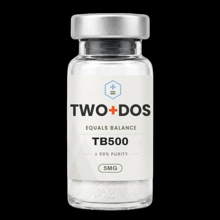
Chemical and physical properties of Thymosin Beta-4 (TB-500)
| Peptide class | Beta-thymosin family. A single WH2-related actin-binding module, intrinsically disordered when free in aqueous buffer and folding only on contact with its partner. |
|---|---|
| Chain length | 43 residues in the mature chain. The UniProt P62328 translated entry lists 44 residues because it retains the initiator methionine that is removed during maturation. |
| Sequence | SDKPDMAEIEKFDKSKLKKTETQEKNPLPSKETIEQEKQAGES (single-letter code, N to C) |
| N-terminal modification | The alpha-amino group of serine-1 carries an acetyl group in the native protein, reported in the original 1981 sequence determination and annotated at UniProt P62328 as N-acetylserine. |
| Molecular formula and mass | C212H350N56O78S, 4963.4 g/mol for the unacetylated 43-residue chain (PubChem CID 45382195). Low and colleagues reported 4982 for the acetylated calf thymus isolate in 1981. |
| CAS registry number | 77591-33-4 |
| PubChem CID | 45382195 |
| Gene and protein database entries | TMSB4X (X-linked), UniProt P62328; the Y-linked paralogue is TMSB4Y |
| Actin-binding motif | LKKTET at residues 17 to 22. The seven-residue span LKKTETQ (17 to 23) is the segment reproduced, in acetylated form, by the separate heptapeptide sold as TB-500. |
| Measured affinity for monomeric actin | 0.32 plus or minus 0.02 micromolar by fluorescence anisotropy (Xue et al., 2014). Weber et al. reported 0.4 to 0.7 micromolar against platelet actin and 2 to 3 micromolar against skeletal muscle actin. Affinity for ADP-loaded actin is roughly two orders of magnitude weaker. |
| Reactive residues | One methionine (position 6) and no cysteine. Disulfide scrambling is therefore not a degradation route for this sequence; oxidation of the single methionine is the principal covalent liability. |
| Physical form, solubility, storage | Lyophilized powder. Aqueous solubility near 50 mg/mL is quoted across chemical catalogues, alongside storage below minus 20 degrees Celsius, protected from light and under inert gas. These are vendor specifications rather than values from the peer-reviewed record. |
What is thymosin beta-4?
Thymosin beta-4 is a 43-amino-acid peptide of the beta-thymosin family, first sequenced from calf thymus in 1981 by Low, Hu and Goldstein. Thymosin beta-4 occurs in the cytoplasm of nearly all nucleated vertebrate cells and is encoded in humans by TMSB4X on the X chromosome, with a Y-linked paralogue designated TMSB4Y.
Its cellular abundance is what distinguishes it from most signalling peptides. Weber and colleagues measured concentrations of 560 micromolar thymosin beta-4 against 280 micromolar monomeric actin inside resting human platelets, meaning the peptide is present at roughly twice the molar amount of the species it binds. At those concentrations it is a bulk buffering component of the cytoskeleton rather than a trace regulator.
The name is a historical artefact. The molecule was isolated from a crude thymic preparation known as fraction 5 and characterized as a thymic hormone, and only later was the same chain shown to be indistinguishable from Fx, the actin-sequestering activity that cell biologists had been tracking independently. Both literatures describe one molecule.
UniProt annotates the human entry with N-acetylserine at position 2 of the translated sequence, phosphothreonine at positions 23 and 34, phosphoserine at positions 2 and 31, and several acetyl-lysine sites. Synthetic and recombinant preparations reproduce the backbone; whether they reproduce the modification pattern depends entirely on the production route.
How does thymosin beta-4 bind G-actin?
Thymosin beta-4 binds a single monomer of G-actin and blocks its addition to a filament. Xue and colleagues measured a dissociation constant of 0.32 plus or minus 0.02 micromolar by fluorescence anisotropy in 2014. Earlier work by Weber and colleagues gave 0.4 to 0.7 micromolar against platelet actin and 2 to 3 micromolar against skeletal muscle actin.
Nucleotide state governs the interaction. Reported constants cluster between 0.1 and 3.9 micromolar for ATP-loaded actin and between 80 and 100 micromolar for ADP-loaded actin, a roughly hundred-fold discrimination. The consequence is a reservoir of polymerization-competent ATP-actin held in reserve, with spent ADP-actin released preferentially.
Two crystal structures published by Xue, Leyrat, Grimes and Robinson in 2014 resolved the geometry, one at 2.3 angstroms using a Pichia actin hybrid and one at 2.0 angstroms using rabbit skeletal muscle actin with a Cobl peptide. Thymosin beta-4 grips the monomer with two short helices: an N-terminal helix spanning aspartate-5 to lysine-11 that contacts the barbed face, and a C-terminal helix from lysine-31 to glutamine-39 that bridges the nucleotide cleft. Total buried interface was about 1,627 square angstroms, of which the C-terminal helix alone contributed 392.
The same structures explain how actin is handed onward to profilin. The C-terminal helix of thymosin beta-4 stabilizes a closed, flattened nucleotide cleft, whereas profilin favours an open cleft, so the two ligands stabilize incompatible conformations. Affinity of thymosin beta-4 for a profilin-actin complex fell about 25-fold relative to free actin, and a ternary intermediate allows transfer without a fully dissociated monomer.
In free solution the peptide has no fixed fold. Czisch and colleagues showed by NMR spectroscopy in 1993 that thymosin beta-4 lacks a uniquely folded conformation in water and carries only transient helical preference; defined helices covering residues 5 to 16 and 31 to 39 appear when the peptide is placed in helix-promoting solvent such as trifluoroethanol. Structure is induced by the binding partner, not carried into the encounter.
Is thymosin beta-4 the same molecule as TB-500?
Thymosin beta-4 and TB-500 are not the same chemical entity, although catalogue listings routinely conflate them. Thymosin beta-4 is the full 43-residue chain of 4963 g/mol. TB-500 material has been analytically identified as the acetylated heptapeptide Ac-LKKTETQ, corresponding to positions 17 through 23 of the parent chain and weighing roughly 889 g/mol, under a fifth of the parent mass.
The distinction matters when reading mechanism papers. Crystallography, anisotropy titrations, the profilin exchange work, and essentially all the cardiac, renal and ocular preclinical literature summarized below used full-length protein, whether synthetic or recombinant. Those measurements describe the 43-residue chain. A seven-residue fragment lacks both terminal helices resolved in the crystal structures, so the actin geometry established for the parent does not transfer to it automatically.
Some published work does bridge the two, having localized angiogenesis-associated activity in specific assays to the actin-binding motif that the short fragment reproduces. That is a narrower claim than equivalence, and the two entities carry different CAS numbers, different formulae and different PubChem records.
What is the Ac-SDKP axis of thymosin beta-4?
Thymosin beta-4 is the physiological precursor of Ac-SDKP, an N-acetylated tetrapeptide corresponding to its own first four residues, serine-aspartate-lysine-proline. Release is a two-enzyme process: meprin-alpha first cuts the 43-residue chain into intermediates shorter than 30 residues, then prolyl oligopeptidase liberates the tetrapeptide, which prolyl oligopeptidase cannot excise from the intact chain directly.
Clearance runs through a well-characterized enzyme. Ac-SDKP is hydrolysed by the N-terminal catalytic domain of angiotensin-converting enzyme, and circulating concentrations of the tetrapeptide rise roughly five-fold when that enzyme is inhibited. This places a fragment of thymosin beta-4 inside a pathway that ordinary cardiovascular pharmacology already perturbs.
Wang, Jia and Zhang reviewed this axis in 2022 across hepatic, renal, cardiac and pulmonary fibrosis models, describing antifibrotic activity attributed to the tetrapeptide in each organ system. For anyone reading thymosin beta-4 papers, the practical implication is that observed effects may belong to the intact chain, to Ac-SDKP, or to both, and few study designs separate them.
What have 2025 and 2026 studies reported about thymosin beta-4?
Recent thymosin beta-4 research has concentrated on ocular surface repair, cardiac ischaemia, and inflammatory injury models, with a parallel strand of protein-engineering work aimed at the peptide's short circulating lifetime. Six studies published between November 2025 and 2026 illustrate the current shape of the field.
In ophthalmology, Nguyen and colleagues reported in Investigative Ophthalmology and Visual Science in 2025 that fusing two copies of the peptide into a tandem construct with two G-actin binding sites raised viability and migration of human telomerase-immortalized corneal epithelial cells above the single-copy peptide, and produced a thicker, continuous epithelial layer in an alkali-injured murine cornea. A 2026 report from Ebrahim and colleagues in the same journal examined adjunctive use alongside ciprofloxacin in Pseudomonas aeruginosa keratitis in C57BL/6 mice, reporting restoration of corneal nerve density toward uninfected control levels.
In cardiac work, Zhang and colleagues published in Cardiovascular Research in 2025 a study pairing a mouse ischaemia-reperfusion model with a randomized, placebo-controlled human trial enrolling 96 people after ST-segment elevation myocardial infarction. In mice, a seven-day course limited cardiac dysfunction and fibrosis assessed 28 days after surgery, with RNA sequencing implicating ErbB2 and Raf1 signalling. In the human arm, infarcted area at 90 days was reduced in the active group, but the overall difference did not reach statistical significance, a result the authors report plainly.
Three 2026 papers extend the inflammatory-injury literature. Ouyang and colleagues reported in Clinical Science that circulating thymosin beta-4 fell in lipopolysaccharide-induced sepsis-associated acute kidney injury and that supplying the peptide suppressed phosphorylation of JNK1/2, p38 MAPK and ERK1/2, with pharmacological MAPK activation partly reversing the effect in HK-2 cells. Ye and colleagues reported in Biomolecules that recombinant human thymosin beta-4 raised survival and lowered circulating pro-inflammatory cytokines in endotoxaemia models, with TLR4 and NF-kappa-B suppression and transcriptomic downregulation of LPAR3. Li and colleagues reported in the Chinese Journal of Contemporary Pediatrics that in BV2 microglial cells challenged with lipopolysaccharide and nigericin, the peptide lowered NLRP3, cleaved caspase-1, GSDMD-N, phospho-STING and phospho-IRF3 protein levels along with mitochondrial reactive oxygen species.
The musculoskeletal evidence is thinner than its popular profile suggests. McGuire, Hughes, Maak and Cushman screened the literature through March 2026 for a scoping review in Applied Sciences and included 80 studies, finding the corpus weighted toward in vitro and mixed designs, concentrated in dermal, vascular and ocular tissue, and sparse in direct musculoskeletal work outside animal models. Biçer and colleagues published one such animal study in 2026, a 32-rat Achilles transection model in which the short fragment group showed significantly higher maximum load to failure at four weeks.
How is research-grade thymosin beta-4 produced and characterized?
Thymosin beta-4 is obtained either by solid-phase peptide synthesis or by recombinant expression in Escherichia coli, and the recombinant route is the one with published process figures. Li and colleagues expressed the peptide in soluble form as a DsbA and hexahistidine fusion, reaching about 50 grams per litre dry cell weight with the fusion at 40 percent of total protein.
Downstream processing in that report used a five-step column sequence combining thermal denaturation, nickel-resin affinity capture and preparative high-performance liquid chromatography, returning material above 98 percent purity with endotoxin, host cell protein and residual DNA inside the limits set by the Chinese Pharmacopoeia. Those are the most concrete purity and impurity numbers in the open literature for this peptide.
Identity confirmation for a 4963 g/mol chain is straightforward by electrospray or quadrupole time-of-flight mass spectrometry after desalting, with reversed-phase C18 chromatography under an acetonitrile gradient used for purity. Two checks matter more than they would for a shorter peptide: whether the N-terminal serine is acetylated, which shifts the observed mass by 42 units, and whether methionine-6 has oxidized, which shifts it by 16.
A recurring theme in the 2025 and 2026 engineering literature is that the native chain does not persist long in circulation. The tandem-construct work cited short circulating lifetime and synthesis cost as the explicit motivation, and a separate 2026 report described a thiol-site-specific PEGylated variant built for the same reason. Both are indirect evidence that the unmodified peptide is cleared rapidly.
Summary of published research
Findings below are reported as published by the cited authors, in the model systems they used. They describe laboratory research, and none of them characterize use in humans.
Low TL, Hu SK, Goldstein AL. Complete amino acid sequence of bovine thymosin beta 4: a thymic hormone that induces terminal deoxynucleotidyl transferase activity in thymocyte populations. Proceedings of the National Academy of Sciences of the United States of America (1981)
- Model system
- Protein chemistry; peptide isolated from calf thymus fraction 5
- Conditions
- Sequential Edman degradation and enzymatic fragmentation of the purified bovine isolate
- Reported finding
- The isolated chain comprised 43 amino acid residues with a reported molecular weight of 4982, and the authors established that the amino terminus is blocked by an acetyl group. This remains the canonical source for the primary structure and the N-terminal acetylation.
Weber A, Nachmias VT, Pennise CR, Pring M, Safer D. Interaction of thymosin beta 4 with muscle and platelet actin: implications for actin sequestration in resting platelets. Biochemistry (1992)
- Model system
- Cell-free biochemistry with purified muscle and platelet actin, plus quantitation in resting human platelets
- Conditions
- Physiological salt; equilibrium binding measurement and direct quantitation of the two species in platelet lysate
- Reported finding
- A 1:1 complex formed with monomeric actin under physiological salt, with the constant unaffected by calcium. Reported affinity was 0.4 to 0.7 micromolar for platelet actin. Concentrations inside resting platelets were measured at 560 micromolar peptide against 280 micromolar monomeric actin. No binding to filament ends or filament sides was detected.
Czisch M, Schleicher M, Hörger S, Voelter W, Holak TA. Conformation of thymosin beta 4 in water determined by NMR spectroscopy. European Journal of Biochemistry (1993)
- Model system
- Cell-free solution-state NMR spectroscopy
- Conditions
- Aqueous buffer, compared against helix-promoting cosolvent conditions
- Reported finding
- The peptide lacked a uniquely folded conformation in water while retaining some preferential alpha-helical population. Defined helices appeared only in helix-promoting solvent, spanning roughly residues 5 to 16 and 31 to 39, identifying the molecule as intrinsically disordered in its free state.
Li T, Ma SY, Tang XC, Sun BG, Tao WM, Sun M. Production and characterization of highly purified recombinant thymosin beta 4 in Escherichia coli. Protein Expression and Purification (2013)
- Model system
- Recombinant expression and downstream process development in Escherichia coli
- Conditions
- Soluble DsbA and hexahistidine fusion; high cell density cultivation; five-step column purification
- Reported finding
- Final biomass reached about 50 grams per litre dry cell weight with the fusion protein at 40 percent expression. Purified material exceeded 98 percent purity, and endotoxin, host cell protein and residual DNA all fell within Chinese Pharmacopoeia limits. Reported as the first highly purified preparation from a genetically engineered source.
Xue B, Leyrat C, Grimes JM, Robinson RC. Structural basis of thymosin-β4/profilin exchange leading to actin filament polymerization. Proceedings of the National Academy of Sciences of the United States of America (2014)
- Model system
- Cell-free structural biology; X-ray crystallography with fluorescence anisotropy binding measurement
- Conditions
- Two complexes solved at 2.3 angstroms (Pichia actin hybrid) and 2.0 angstroms (rabbit skeletal muscle actin with a Cobl peptide)
- Reported finding
- An N-terminal helix from aspartate-5 to lysine-11 contacted the barbed face while a C-terminal helix from lysine-31 to glutamine-39 bridged the nucleotide cleft, burying about 1,627 square angstroms in total with 392 from the C-terminal helix alone. Fluorescence anisotropy gave a dissociation constant of 0.32 plus or minus 0.02 micromolar. Affinity fell roughly 25-fold against a profilin-actin complex.
Nguyen J, Verma S, Vuong VT, Queener H, Coulson-Thomas VJ, Gesteira TF. Engineered Tandem Thymosin Peptide Promotes Corneal Wound Healing. Investigative Ophthalmology and Visual Science (2025)
- Model system
- Human telomerase-immortalized corneal epithelial cells (hTCEpi) plus a murine alkali corneal injury model
- Conditions
- Tandem construct fusing two monomers to create two G-actin binding sites; structural modelling, viability and migration assays, in vivo corneal repair scoring
- Reported finding
- The tandem construct raised corneal epithelial cell viability and migration relative to the single-copy peptide, and modelling indicated simultaneous engagement of two G-actin monomers. In the alkali-injured murine cornea it produced a thicker, continuous epithelial layer approaching unwounded architecture, with reduced scarring compared with the monomer.
Zhang Y, Dong Q, Bian X, et al. Recombinant human thymosin beta 4 improves ischemic cardiac dysfunction in mice and patients with acute ST-segment elevation myocardial infarction after reperfusion. Cardiovascular Research (2025)
- Model system
- C57BL/6J mouse ischaemia-reperfusion model, cardiomyocyte hypoxia-reoxygenation in vitro, and a randomized placebo-controlled human trial with 96 enrolled
- Conditions
- Seven-day course in mice with assessment at 28 days; RNA sequencing; cardiac magnetic resonance imaging in the human arm at 90 days
- Reported finding
- In mice the peptide limited cardiac dysfunction and fibrosis at 28 days, with RNA sequencing implicating ErbB signalling and in vitro work showing ErbB2 and Raf1 activation alongside reduced cardiomyocyte apoptosis. In the human arm, infarcted area at 90-day follow-up was smaller in the active group, but the overall between-group difference was not statistically significant.
Ouyang X, Long Z, Xu X, et al. Thymosin β4 alleviates sepsis-associated acute kidney injury by suppressing MAPK signaling pathway. Clinical Science (2026)
- Model system
- Rodent lipopolysaccharide-induced sepsis-associated acute kidney injury with HK-2 human proximal tubular cells
- Conditions
- Transcriptome profiling, western blotting of MAPK phosphorylation, and pharmacological MAPK activation as a reversal control
- Reported finding
- Circulating peptide concentrations fell significantly in the injury model. Supplying the peptide improved renal function readouts and reduced inflammation and apoptosis, with suppressed phosphorylation of JNK1/2, p38 MAPK and ERK1/2. Pharmacological MAPK activation partially reversed the reduction in pro-inflammatory cytokine expression in HK-2 cells.
Ye Y, Yang X, Liu Y, et al. Recombinant Human Thymosin β4 Attenuates Endotoxemia-Induced ALI and EAE by Suppressing Inflammatory and Oxidative Responses. Biomolecules (2026)
- Model system
- Rodent lipopolysaccharide endotoxaemia models of acute lung injury and encephalopathy
- Conditions
- Survival tracking, cytokine panels, macrophage and microglial profiling, and transcriptomic analysis
- Reported finding
- Recombinant material raised survival rates and reduced systemic overproduction of multiple pro-inflammatory cytokines. Protection was attributed to TLR4 and NF-kappa-B suppression with macrophage modulation, and transcriptomics identified downregulation of LPAR3 as a contributor to the microglial component.
Li YX, Chen CL, Zheng SD, et al. Thymosin β4 inhibits pyroptosis in BV2 microglial cells: a mechanistic study in vitro. Chinese Journal of Contemporary Pediatrics (2026)
- Model system
- BV2 murine microglial cell line
- Conditions
- Lipopolysaccharide plus nigericin co-challenge as an in vitro sepsis-associated encephalopathy model
- Reported finding
- Relative to challenged controls, the peptide reduced mRNA for IL-1beta, IFIT1 and IFN-beta, lowered IL-1beta in the supernatant, and decreased NLRP3, GSDMD-N, cleaved caspase-1, phospho-STING and phospho-IRF3 protein levels, cell death rate and mitochondrial reactive oxygen species, all at P below 0.05. The authors attributed the effect to cGAS-STING pathway regulation.
Biçer O, Adanir O, Güleryüz Y, et al. Effects of BPC-157 and TB-500 on Achilles tendon healing in rats: A histopathological and biomechanical study. Joint Diseases and Related Surgery (2026)
- Model system
- Rodent; 32 male Sprague-Dawley rats with standardized Achilles transection and repair
- Conditions
- Four groups over four weeks; biomechanical load-to-failure testing, Bonar and Movin histopathological scoring, Sirius red collagen analysis
- Reported finding
- The short-fragment group showed significantly higher maximum load to failure at four weeks (P below 0.05), with lower Bonar and Movin scores than controls and increased type I collagen organization. Combining the two peptides conferred no additional advantage over either alone.
McGuire F, Hughes E, Maak T, Cushman DM. Thymosin Beta-4 and TB-500 in Tissue Healing, Regeneration, and Musculoskeletal Repair: A Scoping Review. Applied Sciences (2026)
- Model system
- Review; scoping review of PubMed, Europe PMC and ClinicalTrials.gov records
- Conditions
- Database search executed through March 2026, with 80 records meeting inclusion criteria
- Reported finding
- The included corpus was weighted toward in vitro and mixed designs and concentrated in dermal, vascular and ocular tissue, with most work using the full-length peptide rather than the short fragment. Direct musculoskeletal evidence was limited and human data sparse outside ocular and dermal settings, which the authors framed as popular interest outpacing clinical validation.
What laboratory handling information is published?
Published handling data for thymosin beta-4 is thinner than the volume of biological literature suggests. Chemical catalogues list the material as a lyophilized powder with aqueous solubility around 50 mg/mL, equivalent to roughly 10 millimolar at a formula weight of 4963.4, and recommend storage below minus 20 degrees Celsius in the dark under inert gas. These figures are vendor specifications; no peer-reviewed solubility profile across pH or formal shelf-life study was located.
The sequence itself indicates the chemistry to watch. There is exactly one methionine, at position 6, and no cysteine anywhere in the 43 residues. Disulfide scrambling is therefore not available as a degradation route, and methionine sulfoxide formation is the principal covalent liability to monitor, detectable as a 16-unit mass shift by electrospray or quadrupole time-of-flight mass spectrometry.
Two identity checks are worth building into any incoming-material protocol. The first is acetylation state at the N-terminal serine, which separates the native modified chain from an unacetylated synthetic one by 42 mass units. The second is whether the material is the 43-residue chain at all rather than the acetylated heptapeptide, since the two differ by more than 4,000 g/mol and are trivially separated by mass spectrometry despite sharing catalogue names.
Because the peptide is intrinsically disordered when free, circular dichroism of the material in plain aqueous buffer is expected to read as predominantly random coil, and a strongly helical spectrum in water would itself be an anomaly worth investigating. Recombinant preparations reported above 98 percent purity by the five-step process cited here, which is a reasonable published benchmark for comparison against a certificate of analysis.
Frequently asked research questions
What molecular target does thymosin beta-4 have?
Monomeric G-actin, in a 1:1 complex. This is one of the better-quantified interactions in the peptide literature: a dissociation constant of 0.32 plus or minus 0.02 micromolar by fluorescence anisotropy, two crystal structures at 2.3 and 2.0 angstroms, and roughly hundred-fold selectivity for ATP-loaded over ADP-loaded actin.
How large is thymosin beta-4 and what is its formula?
The mature chain is 43 amino acids with the formula C212H350N56O78S and a mass of 4963.4 g/mol for the unacetylated form, catalogued as PubChem CID 45382195 and CAS 77591-33-4. The 1981 characterization of the acetylated calf thymus isolate reported 4982.
Does thymosin beta-4 have a fixed three-dimensional structure?
Not on its own. NMR spectroscopy in water showed no uniquely folded conformation, only transient helical preference. Two helices covering roughly residues 5 to 16 and 31 to 39 form in helix-promoting solvent, and equivalent helices appear in the crystal structures once actin is bound. The fold is induced by the partner.
Why does thymosin beta-4 appear in fibrosis literature?
Because it is the precursor of Ac-SDKP. Meprin-alpha cleaves the 43-residue chain into shorter intermediates and prolyl oligopeptidase then releases the N-terminal tetrapeptide, which angiotensin-converting enzyme clears. A 2022 review traced antifibrotic activity for that fragment across hepatic, renal, cardiac and pulmonary models, so effects reported for the parent chain may belong to either species.
What is the regulatory and evidence status of thymosin beta-4?
No regulatory authority has cleared thymosin beta-4 as a finished medicine, and a 2026 scoping review of 80 records found the evidence weighted toward in vitro and animal designs with sparse human data outside ocular and dermal settings. The World Anti-Doping Agency lists the compound and its derivatives on its 2026 Prohibited List. Material is supplied for laboratory research only.
Thymosin Beta-4 (TB-500) at TWO+DOS
TWO+DOS supplies Thymosin Beta-4 (TB-500) as a research-use-only compound, third-party tested, with certificates of analysis emailed immediately on request.
View the Thymosin Beta 4 (TB500)listing →Related research overviews
References
- PubChem CID 45382195: thymosin beta-4 formula, mass and identifier record
- UniProt P62328 (TMSB4X): sequence, modifications and function annotation
- Low, Hu and Goldstein 1981, PNAS: complete amino acid sequence (PMID 6940133)
- Weber et al. 1992, Biochemistry: actin affinity and platelet concentrations (PMID 1627561)
- Czisch et al. 1993, European Journal of Biochemistry: solution NMR (PMID 8269922)
- Xue et al. 2014, PNAS: crystal structures and profilin exchange (PMID 25313062)
- Li et al. 2013, Protein Expression and Purification: recombinant production (PMID 23711379)
- Nguyen et al. 2025, Investigative Ophthalmology and Visual Science (PMID 41235866)
- Zhang et al. 2025, Cardiovascular Research (PMID 41229390)
- Ouyang et al. 2026, Clinical Science (PMID 42417058)
- Ye et al. 2026, Biomolecules (PMID 42352234)
- Wang, Jia and Zhang 2022, IJMS: the Tbeta4-POP-Ac-SDKP axis (PMID 36362069)
- McGuire et al. 2026, Applied Sciences: scoping review of 80 records
For research use only. Not for human or veterinary use. Not for diagnostic or therapeutic use.