
KPV (Lys-Pro-Val): PepT1 Transport and NF-κB Signaling Research
By the TWO+DOS Research Team · Published 2026-08-13
For research use only. Not for human or veterinary use. Not for diagnostic or therapeutic use.
KPV is a synthetic tripeptide composed of lysine, proline and valine, corresponding to residues 11 to 13 at the C-terminus of α-melanocyte-stimulating hormone. Published research examines it as a small, highly polar molecule that enters cells through the proton-coupled peptide transporter PepT1 and dampens NF-κB and MAP kinase signaling.
This overview gathers what peer-reviewed sources report for the molecule: verified chemical identifiers and computed physicochemical values, the transporter biology that governs where the fragment accumulates, the signaling readouts that recur across the literature, the question of whether melanocortin receptors are involved at all, and findings from cell-line, rodent and formulation studies published between 2003 and 2026. All material described here is for laboratory research use only.
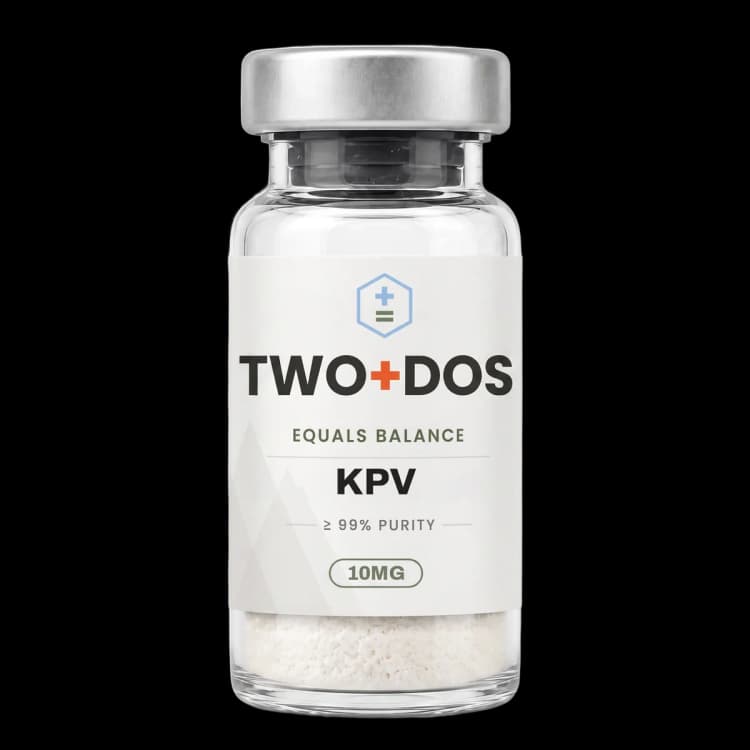
Chemical and physical properties of KPV
| Compound class | Linear tripeptide; C-terminal fragment of α-melanocyte-stimulating hormone |
|---|---|
| Residue sequence | Lys-Pro-Val, corresponding to residues 11, 12 and 13 of the 13-residue parent hormone |
| Parent hormone sequence | Ac-Ser-Tyr-Ser-Met-Glu-His-Phe-Arg-Trp-Gly-Lys-Pro-Val-NH2 |
| Monoisotopic mass | 342.2267 Da (PubChem CID 125672) |
| Computed XLogP | -3.5 (PubChem), describing a strongly hydrophilic molecule |
| Topological polar surface area | 139 Ų (PubChem computed) |
| Rotatable bonds | 9 (PubChem computed) |
| Hydrogen bonding | 4 donors and 6 acceptors (PubChem computed) |
| Reported transport route | PepT1 (SLC15A1), the proton-coupled di- and tripeptide transporter |
| Absent pharmacophore | Lacks the His-Phe-Arg-Trp core (α-MSH residues 6-9) recognized by melanocortin receptors |
| Reported degradation product | Lys-Pro-diketopiperazine under acid, alkali and peroxide stress |
What is KPV and where does it sit in the α-MSH sequence?
KPV is the C-terminal tripeptide of α-melanocyte-stimulating hormone, a 13-residue neuropeptide with the sequence Ac-Ser-Tyr-Ser-Met-Glu-His-Phe-Arg-Trp-Gly-Lys-Pro-Val-NH2. Lysine, proline and valine occupy positions 11, 12 and 13. PubChem records the fragment under CID 125672 with the formula C16H30N4O4 and a molecular weight of 342.43 g/mol.
Three residues is unusually small for a research peptide, and the computed properties reflect that. PubChem lists a monoisotopic mass of 342.2267 Da, an XLogP of -3.5, a topological polar surface area of 139 Ų, nine rotatable bonds, four hydrogen bond donors and six acceptors. Those values describe a compact, strongly hydrophilic solid rather than a membrane-partitioning molecule, which is why transporter biology rather than passive diffusion dominates the mechanistic literature.
The lysine side chain carries a positive charge at neutral pH and the C-terminal carboxylate carries a negative one, so the free acid form is zwitterionic. Proline in the central position constrains the backbone through its pyrrolidine ring, limiting conformational freedom across the middle of the molecule. Some suppliers list an acetylated, amidated variant, Ac-KPV-NH2, which is a chemically distinct entity from the free acid recorded under CAS 67727-97-3.
Naming across the literature is inconsistent enough to cause confusion. The same molecule appears as KPV, Lys-Pro-Val, α-MSH (11-13), MSH (11-13) and tripeptide KPV. A related but separate fragment, KdPT, substitutes D-proline and threonine and belongs to a different line of work; the two are frequently discussed side by side in review articles and should not be conflated.
How does PepT1 transport bring KPV into cells?
KPV enters cells through PepT1, the proton-coupled di- and tripeptide transporter encoded by SLC15A1. Dalmasso and colleagues measured uptake directly in 2008 using tritiated KPV alongside competition with unlabeled peptide, reporting transporter-mediated accumulation in human intestinal epithelial cells and in T cells rather than nonspecific entry across the plasma membrane.
The distribution of PepT1 is what makes this route interesting to the intestinal literature. The transporter is abundant in small intestine under normal conditions and is induced in colonic tissue during inflammatory bowel disease, and it is also expressed by immune cells. A tripeptide that depends on PepT1 therefore concentrates preferentially in exactly the tissue compartments where the transporter is upregulated, which supplies a mechanism for tissue selectivity without any targeting ligand attached to the molecule.
Transporter dependence has been probed genetically as well as pharmacologically. Viennois and colleagues reported in 2016 that transgenic mice overexpressing human PepT1 developed larger tumors, greater tumor burden and increased intestinal inflammation in a colitis-associated cancer model, while PepT1 knockout mice showed significantly decreased tumor number, tumor size and intestinal inflammation. KPV delivered to wild-type animals limited carcinogenesis in the same model through a PepT1-dependent route.
Which signaling pathways do KPV studies measure?
KPV research centers on two signaling readouts: nuclear factor kappa B and the mitogen-activated protein kinase cascade. Dalmasso and colleagues reported that nanomolar concentrations of the tripeptide inhibited activation of both pathways in cytokine-stimulated human intestinal epithelial cells and T cells, and reduced secretion of pro-inflammatory cytokines.
Where in the NF-κB pathway the fragment acts has been addressed separately. Land reported in 2012 that KPV and γ-MSH each produced concentration-dependent inhibition of NF-κB signaling, matrix metalloproteinase-9 activity, and IL-8 and eotaxin secretion in human bronchial epithelial cells challenged with TNF-α or respiratory syncytial virus. The two peptides diverged mechanistically: KPV blocked nuclear import of the p65RelA subunit, whereas γ-MSH acted through MC3R.
MAP kinase branches recur in the more recent cell work. Sung and colleagues reported in 2025 that KPV limited reactive oxygen species production in HaCaT keratinocytes, and that the reduction in oxidant signal was upstream of extracellular signal-regulated kinase and p38 activation. The same study tracked NF-κB as a redox-sensitive transcription factor and reported reduced expression of Bax, Bcl-2 and cleaved caspase-3.
Inflammasome output appears as a third readout. In the keratinocyte work, KPV blocked reactive oxygen species-mediated caspase-1 activation and lowered IL-1β secretion, a sequence the authors describe as mitigation of pyroptosis. Getting and colleagues had earlier reported IL-1β inhibition as a candidate route in a rodent crystal-induced peritonitis model, so the cytokine recurs across two decades and two very different systems.
Does KPV activity depend on melanocortin receptors?
KPV lacks the His-Phe-Arg-Trp core that gives α-MSH its affinity for melanocortin receptors. That message sequence occupies residues 6 to 9 of the parent hormone, while KPV is drawn from residues 11 to 13, so the fragment carries none of the pharmacophore that MC1R through MC5R recognize. The structural argument is straightforward, and two experimental lines support it.
The first is genetic. Kannengiesser and colleagues ran DSS colitis in mice carrying a nonfunctional melanocortin-1 receptor, the MC1Re/e strain, and reported that KPV rescued all animals in the peptide arm from death during the challenge. Activity in an animal lacking functional MC1R indicates a mechanism at least partially independent of that receptor.
The second is biochemical. Getting and colleagues compared KPV against α-MSH and the melanocortin agonist MTII in crystal-induced peritonitis. All three reduced polymorphonuclear leukocyte accumulation in the peritoneal cavity, but MTII raised cyclic AMP while KPV did not, and the authors concluded that KPV is unlikely to act through melanocortin receptors.
This matters for how the compound is positioned in reviews. Gravina and colleagues surveyed the melanocortin system in inflammatory bowel disease in 2023 and describe a family of mediators and receptors spanning melanogenesis, steroidogenesis and neuromodulation. KPV occupies an odd position inside that family: derived from the prototypical melanocortin agonist, yet apparently operating through transporter-mediated uptake and intracellular signaling rather than receptor occupancy.
What have 2023 to 2026 studies reported about KPV?
KPV appears in the 2023 to 2026 literature across three model families: colonic inflammation in rodents, keratinocyte and reconstructed skin models challenged with particulate matter, and metabolic cell lines. Working concentrations in the recent cell studies cluster at 50 to 100 µg/mL, far above the nanomolar range reported for NF-κB inhibition in intestinal epithelium.
The 2025 keratinocyte study supplies the most detailed cell-model numbers. PM10 particulate matter suppressed HaCaT proliferation and raised IL-1β secretion; KPV at 50 µg/mL restored viability and lowered IL-1β, acting through reduced oxidant production, blunted ERK and p38 activation, and suppressed NF-κB. A three-dimensional reconstructed skin model showed attenuated inflammatory cell death under the same challenge.
Two 2026 papers from overlapping Korean groups moved the fragment into lipid-handling cell models. In HepG2 hepatic epithelial cells challenged with oleic acid, KPV at 100 µg/mL lowered lipid accumulation and fatty acid synthase expression without cytotoxicity, with reduced reactive oxygen species, blunted ERK activation, lower AKT and mTORC1 phosphorylation, and altered PPARγ phosphorylation. In 3T3-L1 preadipocytes under MDI differentiation, KPV at 100 µg/mL reduced Oil Red O staining intensity by roughly 55% and intracellular triglyceride content by roughly 38% against the MDI comparator, again with lower PPARγ and fatty acid synthase expression.
Delivery chemistry produced the newest intestinal result. Cheng and colleagues published a self-immolative conjugate platform in Science Advances in 2026 in which the linker is cleaved by the inflammatory environment itself. The KPV-based conjugate, designated proKPV, reached 3.8-fold greater colonic accumulation than the free tripeptide in colitis models while retaining activity at a lower delivered amount.
Review coverage has kept pace. Adnan and colleagues surveyed tripeptides in skin repair across studies published from 2016 to 2025, and record KPV-loaded hydrogels as reducing inflammation, supporting tissue regeneration and acting against methicillin-resistant Staphylococcus aureus.
How has KPV been formulated in published delivery research?
KPV formulation research responds to a practical problem: a three-residue peptide clears rapidly and distributes broadly, so most published work encapsulates or conjugates it. Xiao and colleagues loaded the tripeptide into hyaluronic acid-functionalized polymeric nanoparticles of roughly 272.3 nm with a zeta potential near -5.3 mV, then embedded those particles in a chitosan and alginate hydrogel.
That construct was reported to release nanoparticles in the colonic lumen, penetrate colitis tissue, and internalize into colonic epithelial cells and macrophages, showing greater capacity to limit mucosal damage and lower TNF-α than the unfunctionalized comparator. Marotti and colleagues extended the surface-design logic in 2023 with hybrid lipid hyaluronate-KPV conjugated nanoparticles carrying teduglutide, reporting that immunomodulation depended on whether the hyaluronan-KPV functionalization was present.
Carrier-free approaches have also been described. Zhang and colleagues reported in 2024 that KPV and rapamycin self-assemble into nanoparticles without an added carrier material, and that the resulting construct inhibited vascular calcification in cell and rodent systems through combined suppression of inflammatory signaling and activation of autophagy. A separate 2024 report co-assembled KPV with the immunosuppressant FK506 into a PepT1-targeted nanodrug evaluated against acute and chronic DSS colitis.
Skin delivery has been studied physically rather than chemically. Pawar and colleagues measured transdermal transport of KPV across microporated human skin, reporting permeation of 4.4 µg/cm² per hour with microneedle poration alone and roughly 35-fold enhancement when iontophoresis was combined with poration, with confocal imaging confirming migration beyond 100 µm of depth.
Summary of published research
Findings below are reported as published by the cited authors, in the model systems they used. They describe laboratory research, and none of them characterize use in humans.
Getting SJ, Schiöth HB, Perretti M. Dissection of the anti-inflammatory effect of the core and C-terminal (KPV) alpha-melanocyte-stimulating hormone peptides. Journal of Pharmacology and Experimental Therapeutics (2003)
- Model system
- rodent (murine crystal-induced peritonitis)
- Conditions
- systemic delivery of KPV, α-MSH or the melanocortin agonist MTII; peritoneal leukocyte counts and cyclic AMP measurement
- Reported finding
- KPV, α-MSH and MTII each produced a significant reduction in polymorphonuclear leukocyte accumulation in the peritoneal cavity, but MTII raised cyclic AMP while KPV did not; the authors concluded KPV is unlikely to mediate its effects through melanocortin receptors and identified IL-1β inhibition as a candidate route.
Dalmasso G, Charrier-Hisamuddin L, Nguyen HT, Yan Y, Sitaraman S, Merlin D. PepT1-mediated tripeptide KPV uptake reduces intestinal inflammation. Gastroenterology (2008)
- Model system
- human cell lines (intestinal epithelial cells and T cells) plus rodent colitis models
- Conditions
- cytokine stimulation with and without KPV; NF-κB luciferase reporter, Western blot, RT-PCR and ELISA; uptake with unlabeled and tritiated KPV; KPV supplied in drinking water in DSS- and TNBS-induced colitis
- Reported finding
- Nanomolar concentrations of KPV inhibited activation of NF-κB and MAP kinase signaling and reduced pro-inflammatory cytokine secretion, with uptake proceeding through PepT1 in both immune and epithelial cells; oral delivery reduced the incidence of DSS- and TNBS-induced colitis with decreased pro-inflammatory cytokine expression.
Kannengiesser K, Maaser C, Heidemann J, Luegering A, Ross M, Brzoska T, Bohm M, Luger TA, Domschke W, Kucharzik T. Melanocortin-derived tripeptide KPV has anti-inflammatory potential in murine models of inflammatory bowel disease. Inflammatory Bowel Diseases (2008)
- Model system
- rodent (DSS colitis, CD45RBhi transfer colitis, and MC1Re/e mice)
- Conditions
- DSS and transfer colitis models scored by histology and colonic myeloperoxidase activity; parallel arm in animals carrying a nonfunctional melanocortin-1 receptor
- Reported finding
- Inflammatory infiltrates decreased significantly and colonic myeloperoxidase activity fell in the KPV arm of both colitis models, and in MC1Re/e mice KPV rescued all animals in that arm from death during DSS colitis, which the authors read as a mechanism at least partially independent of MC1R signaling.
An SH, Park JY, Lee SJ. KPV attenuates adipogenesis and lipid metabolism through modulation of ROS-mediated AKT/mTORC1/PPARgamma signaling. Tissue and Cell (2026)
- Model system
- cell line (3T3-L1 preadipocytes)
- Conditions
- MDI-induced adipocyte differentiation; KPV up to 100 µg/mL; Oil Red O staining, intracellular triglyceride quantification and marker expression
- Reported finding
- KPV at 100 µg/mL reduced Oil Red O staining intensity by approximately 55% and intracellular triglyceride content by approximately 38% relative to the MDI comparator, lowered expression of PPARγ and fatty acid synthase, and attenuated reactive oxygen species production alongside reduced AKT-dependent mTOR signaling and altered PPARγ phosphorylation.
Xiao B, Xu Z, Viennois E, Zhang Y, Zhang Z, Zhang M, Han MK, Kang Y, Merlin D. Orally Targeted Delivery of Tripeptide KPV via Hyaluronic Acid-Functionalized Nanoparticles Efficiently Alleviates Ulcerative Colitis. Molecular Therapy (2017)
- Model system
- rodent (murine ulcerative colitis) plus colonic epithelial cells and macrophages
- Conditions
- KPV loaded into hyaluronic acid-functionalized polymeric nanoparticles, delivered orally within a chitosan and alginate hydrogel
- Reported finding
- The nanoparticles measured approximately 272.3 nm with a zeta potential near -5.3 mV, released in the colonic lumen, penetrated colitis tissue and internalized into colonic epithelial cells and macrophages, and showed greater capacity to limit mucosal damage and downregulate TNF-α than the unencapsulated comparator system.
Viennois E, Ingersoll SA, Ayyadurai S, Zhao Y, Wang L, Zhang M, Han MK, Garg P, Xiao B, Merlin D. Critical role of PepT1 in promoting colitis-associated cancer and therapeutic benefits of the anti-inflammatory PepT1-mediated tripeptide KPV in a murine model. Cellular and Molecular Gastroenterology and Hepatology (2016)
- Model system
- rodent (hPepT1 transgenic, PepT1 knockout and wild-type mice in a colitis-associated cancer model)
- Conditions
- comparison of tumor number, tumor size and intestinal inflammation across PepT1 genotypes, with KPV delivered to wild-type animals
- Reported finding
- Transgenic mice overexpressing human PepT1 showed larger tumor sizes, increased tumor burden and increased intestinal inflammation, while tumor number, tumor size and intestinal inflammation were significantly decreased in PepT1 knockout mice; KPV limited carcinogenesis in wild-type animals through a PepT1-dependent route.
Gravina AG, Pellegrino R, Durante T, Palladino G, Imperio G, D'Amico G, Trotta MC, Dallio M, Romeo M, D'Amico M, Federico A. The Melanocortin System in Inflammatory Bowel Diseases: Insights into Its Mechanisms and Therapeutic Potentials. Cells (2023)
- Model system
- review
- Conditions
- narrative synthesis of melanocortin mediator and receptor literature, weighted toward murine colitis models
- Reported finding
- The review described the melanocortin system as a set of mediators and receptors spanning melanogenesis, steroidogenesis, neuromodulation and inflammatory modulation, and summarized evidence that the system counteracts colitis inflammation and addresses cytokine imbalance in damaged intestinal microenvironments.
Marotti V, Xu Y, Bohns Michalowski C, Zhang W, Domingues I, Ameraoui H, Moreels TG, Baatsen P, Van Hul M, Muccioli GG, Cani PD, Alhouayek M, Malfanti A, Beloqui A. A nanoparticle platform for combined mucosal healing and immunomodulation in inflammatory bowel disease treatment. Bioactive Materials (2023)
- Model system
- rodent inflammatory bowel disease model with cell-based immunomodulation assays
- Conditions
- hybrid lipid hyaluronate-KPV conjugated nanoparticles loaded with teduglutide, compared against nanocarriers lacking the hyaluronan-KPV surface
- Reported finding
- Immunosuppressive activity of the nanocarriers depended on the presence or absence of hyaluronan-KPV functionalization, and the platform combined induction of glucagon-like peptide 2 secretion with surface-driven immunomodulation in the same construct.
Zhang D, Jiang L, Yu F, Yan P, Liu Y, Wu Y, Yang X. PepT1-targeted nanodrug based on co-assembly of anti-inflammatory peptide and immunosuppressant for combined treatment of acute and chronic DSS-induced ColitiS. Frontiers in Pharmacology (2024)
- Model system
- rodent (murine acute and chronic DSS colitis)
- Conditions
- five groups receiving saline, 2.5% or 4% DSS, KPV alone, FK506 alone, or the co-assembled nanoparticle
- Reported finding
- The nanoparticle group showed greater improvement in colon length and disease activity index than KPV or FK506 alone, with decreased myeloperoxidase, nitric oxide and reactive oxygen species, decreased TNF-α, IL-1β and IL-6, reduced CD68 and CD3 expression, and restored Claudin-5, Occludin-1 and ZO-1 tight junction proteins.
Zhang L, Li D, Aierken Y, Zhang J, Liu Z, Lin Z, Jiang L, Li Q, Wu Y, Liu Y. KPV and RAPA Self-Assembled into Carrier-Free Nanodrugs for Vascular Calcification Therapy. Advanced Healthcare Materials (2024)
- Model system
- cell culture and rodent vascular calcification models
- Conditions
- carrier-free nanoparticles formed by self-assembly of KPV with rapamycin, an autophagy activator, evaluated against comparator controls
- Reported finding
- The self-assembled KPV and rapamycin nanoparticles showed good stability and inhibited vascular calcification in both cell and animal systems relative to comparator controls, which the authors attributed to combined suppression of the inflammatory response and activation of autophagy.
Sung J, Ju SY, Park S, Jung WK, Je JY, Lee SJ. Lysine-Proline-Valine peptide mitigates fine dust-induced keratinocyte apoptosis and inflammation by regulating oxidative stress and modulating the MAPK/NF-κB pathway. Tissue and Cell (2025)
- Model system
- human cell line (HaCaT keratinocytes) plus a three-dimensional reconstructed skin model
- Conditions
- PM10 particulate matter challenge; KPV at 50 µg/mL; viability, IL-1β ELISA, reactive oxygen species measurement and Western blot
- Reported finding
- PM10 suppressed HaCaT proliferation and raised IL-1β secretion, and KPV at 50 µg/mL restored viability and lowered IL-1β; the peptide inhibited reactive oxygen species production upstream of ERK and p38 activation, decreased Bax, Bcl-2 and cleaved caspase-3, suppressed NF-κB, blocked caspase-1 activation, and attenuated inflammatory cell death in the three-dimensional model.
Cheng J, Wu P, Li C, Han Y, Sun M, Dou Y, Chen S, Zhang J. Inflammation-triggered self-immolative conjugates enable oral peptide delivery by overcoming gastrointestinal barriers. Science Advances (2026)
- Model system
- rodent colitis model with oral delivery
- Conditions
- self-immolative conjugate linker cleaved by the inflammatory microenvironment; the KPV-based construct designated proKPV compared against the free tripeptide
- Reported finding
- The proKPV conjugate reached 3.8-fold greater colonic accumulation than free KPV while retaining activity at a substantially lower delivered amount, which the authors present as evidence that inflammation-triggered self-immolative chemistry can carry a small peptide past gastrointestinal barriers.
What laboratory handling information is published?
KPV is supplied as a lyophilized powder, and its computed profile matches that format. An XLogP of -3.5, a topological polar surface area of 139 Ų and a zwitterionic free-acid form describe a highly water-soluble solid with no lipophilic character. Published methods prepare working solutions in aqueous buffer, saline or culture medium.
Working concentrations in the literature span roughly six orders of magnitude by model type, so a concentration quoted without its model system is close to meaningless. Signaling inhibition in cytokine-stimulated intestinal epithelial cells and T cells was reported at nanomolar concentrations, while recent keratinocyte, hepatocyte and preadipocyte work used 50 to 100 µg/mL, equivalent to roughly 146 to 292 µM at a molecular weight of 342.43 g/mol.
Stability is unusually well characterized for a compound of this size. A validated reversed-phase HPLC method on a C18 column, 4.6 × 250 mm with 5 µm silica, running a gradient of 0.1% trifluoroacetic acid in water against 0.1% trifluoroacetic acid in acetonitrile, achieved a correlation coefficient of 0.9999 with limits of detection and quantification at 0.01 and 0.25 µg/mL. Forced degradation under acid, alkali and hydrogen peroxide produced Lys-Pro-diketopiperazine as the major breakdown product, formed by intramolecular cyclization of the lysine and proline residues, and the method resolves that product from intact peptide.
Identity confirmation follows standard peptide practice: electrospray mass spectrometry against a monoisotopic mass of 342.2267 Da, giving a singly protonated ion near m/z 343.2, with purity determined by reversed-phase HPLC and reported on the lot certificate of analysis. Lyophilized material is normally kept frozen, protected from light and moisture, with aqueous stock solutions aliquoted so that repeated freeze-thaw cycles are avoided. Because the diketopiperazine pathway is pH sensitive, aqueous stocks held at extremes of pH are the condition most likely to degrade.
This material is offered for laboratory research use only. It is not a drug, not a food, and not intended for human or veterinary use.
Frequently asked research questions
What does the abbreviation KPV stand for?
KPV is the single-letter code for the amino acids lysine (K), proline (P) and valine (V), read in that order from the N-terminus. The same molecule appears in the literature as Lys-Pro-Val, α-MSH (11-13), MSH (11-13) and tripeptide KPV, recorded under CAS 67727-97-3 and PubChem CID 125672.
How is KPV related to α-melanocyte-stimulating hormone?
KPV is the last three residues of α-MSH, a 13-residue neuropeptide with the sequence Ac-Ser-Tyr-Ser-Met-Glu-His-Phe-Arg-Trp-Gly-Lys-Pro-Val-NH2. The fragment does not include the His-Phe-Arg-Trp message sequence at positions 6 to 9 that confers melanocortin receptor affinity, so the two molecules are studied as chemically related but mechanistically distinct entities.
Why does the PepT1 transporter matter in KPV research?
PepT1, encoded by SLC15A1, is a proton-coupled transporter that carries di- and tripeptides across cell membranes. It is abundant in small intestine, induced in colonic tissue during inflammatory bowel disease, and expressed by immune cells. Because KPV depends on this route rather than passive diffusion, it accumulates preferentially in tissue where the transporter is upregulated.
What concentrations of KPV appear in published cell studies?
The range is wide and model-dependent. NF-κB and MAP kinase inhibition in cytokine-stimulated intestinal epithelial cells and T cells was reported at nanomolar concentrations, whereas 2025 and 2026 keratinocyte, HepG2 and 3T3-L1 studies used 50 to 100 µg/mL, roughly 146 to 292 µM. Comparing figures across those papers without noting the model system is misleading.
Which model systems dominate the KPV literature?
Four recur across the citations above: murine colitis models induced with DSS or TNBS, human cell lines including intestinal epithelium, HaCaT keratinocytes, HepG2 and 3T3-L1, cell-free analytical and formulation characterization, and nanoparticle or hydrogel delivery systems evaluated in rodents. No completed human trial of the single tripeptide appears in the indexed literature as of August 2026.
KPV at TWO+DOS
TWO+DOS supplies KPV as a research-use-only compound, third-party tested, with certificates of analysis emailed immediately on request.
View the KPV (10mg)listing →Related research overviews
References
- PubChem CID 125672: MSH (11-13) compound summary
- PepT1-mediated tripeptide KPV uptake reduces intestinal inflammation (Gastroenterology 2008)
- Melanocortin-derived tripeptide KPV has anti-inflammatory potential in murine models of inflammatory bowel disease (Inflamm Bowel Dis 2008)
- Dissection of the anti-inflammatory effect of the core and C-terminal (KPV) alpha-melanocyte-stimulating hormone peptides (J Pharmacol Exp Ther 2003)
- Inhibition of cellular and systemic inflammation cues in human bronchial epithelial cells by melanocortin-related peptides (Int J Physiol Pathophysiol Pharmacol 2012)
- Critical role of PepT1 in promoting colitis-associated cancer and therapeutic benefits of the anti-inflammatory PepT1-mediated tripeptide KPV in a murine model (Cell Mol Gastroenterol Hepatol 2016)
- Stability-indicating HPLC assay for lysine-proline-valine (KPV) in aqueous solutions and skin homogenates (Biomed Chromatogr 2015)
- Orally Targeted Delivery of Tripeptide KPV via Hyaluronic Acid-Functionalized Nanoparticles Efficiently Alleviates Ulcerative Colitis (Mol Ther 2017)
- The Melanocortin System in Inflammatory Bowel Diseases: Insights into Its Mechanisms and Therapeutic Potentials (Cells 2023)
- Lysine-Proline-Valine peptide mitigates fine dust-induced keratinocyte apoptosis and inflammation (Tissue Cell 2025)
- Exploring the Role of Tripeptides in Wound Healing and Skin Regeneration: A Comprehensive Review (Int J Med Sci 2025)
- Inflammation-triggered self-immolative conjugates enable oral peptide delivery by overcoming gastrointestinal barriers (Sci Adv 2026)
For research use only. Not for human or veterinary use. Not for diagnostic or therapeutic use.