
Thymosin Alpha-1 (Tα1): Structure, Prothymosin Origin and TLR Signaling Research
By the TWO+DOS Research Team · Published 2026-08-13
For research use only. Not for human or veterinary use. Not for diagnostic or therapeutic use.
Thymosin alpha-1 is a 28-residue, N-terminally acetylated acidic polypeptide corresponding to the first 28 amino acids of the nuclear protein prothymosin alpha. Published under the international nonproprietary name thymalfasin and CAS 62304-98-7, the molecule is studied as an endogenous modulator of dendritic cell and T-cell signaling in immunology laboratories.
This overview collects what peer-reviewed sources report for the peptide: verified chemical identifiers and computed properties, the proteolytic route by which it is released from its parent protein, the conformational change it undergoes at anionic membranes, the Toll-like receptor pathways attributed to it in antigen-presenting cells, and results from cell-line, rodent and controlled human studies published between 1977 and 2026. All material described here is for laboratory research use only.
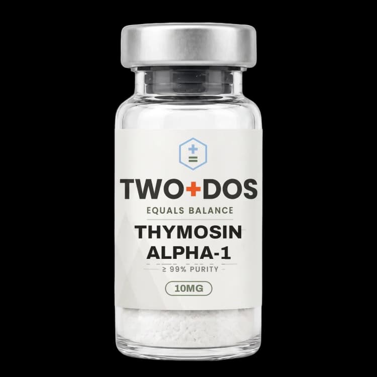
Chemical and physical properties of Thymosin Alpha-1
| Compound class | 28-residue Nα-acetylated acidic polypeptide |
|---|---|
| Chain composition | 28 amino acids, no cysteine and therefore no disulfide bond |
| N-terminus | Acetylated serine; the α-amino group is blocked, not free |
| C-terminus | Free carboxylic acid at asparagine 28 |
| Parent protein | Residues 1-28 of prothymosin alpha, a 109-residue nuclear protein |
| Monoisotopic mass | 3106.5041 Da (PubChem CID 16130571) |
| Exact mass | 3107.5075 Da (PubChem computed) |
| Side-chain charge balance | 6 glutamate and 3 aspartate residues against 4 lysine residues; no arginine, no histidine |
| Isoelectric point | Reported near pH 4.0-4.3 by slab gel isoelectric focusing |
| Computed XLogP | -24 (PubChem), an extreme hydrophilic value for a peptide of this size |
| Topological polar surface area | 1460 Ų, with 49 hydrogen bond donors and 59 acceptors (PubChem computed) |
What is the primary structure of the thymosin alpha-1 peptide?
Thymosin alpha-1 carries the sequence Ac-Ser-Asp-Ala-Ala-Val-Asp-Thr-Ser-Ser-Glu-Ile-Thr-Thr-Lys-Asp-Leu-Lys-Glu-Lys-Lys-Glu-Val-Val-Glu-Glu-Ala-Glu-Asn, an acidic 28-residue chain that contains no cysteine and therefore forms no disulfide bond. PubChem CID 16130571 records the molecular formula C129H215N33O55, a molecular weight of 3108.3 g/mol and a monoisotopic mass of 3106.5041 Da.
Composition explains the peptide's behaviour in buffer. The chain carries six glutamate and three aspartate residues against only four lysines, with no arginine and no histidine, so at neutral pH the nine acidic side chains and the C-terminal carboxylate outweigh the basic ones. The N-terminal acetyl group removes the positive charge a free α-amino terminus would supply, pushing the net charge further negative and matching the isoelectric point reported near pH 4.0 to 4.3 by isoelectric focusing.
An unusual feature of the sequence is what it lacks. Thymosin alpha-1 contains no tryptophan, no tyrosine and no phenylalanine, so the molecule has no aromatic chromophore. PubChem computes an XLogP of -24, a topological polar surface area of 1460 Ų, 49 hydrogen bond donors, 59 acceptors and 111 rotatable bonds, describing a highly solvated, conformationally flexible chain with no lipophilic character and no fixed shape to preserve.
How is thymosin alpha-1 generated from prothymosin alpha?
Thymosin alpha-1 arises inside cells as a proteolytic fragment rather than as an independent gene product. Sarandeses, Covelo, Díaz-Jullien and Freire reported in the Journal of Biological Chemistry in 2003 that mammalian legumain, a lysosomal asparaginyl endopeptidase, cleaves prothymosin alpha and releases the peptide from the parent chain.
The cleavage chemistry is specific. Legumain hydrolyses asparaginyl bonds, and in prothymosin alpha the enzyme was reported to cut at Asn28-Gly29 and at Asn35-Gly36 with comparable in-vitro efficiency. The first cut yields the 28-residue thymosin alpha-1; the second yields the 35-residue thymosin alpha-11. Both share the same acetylated N-terminus and differ only in how far the chain extends.
Prothymosin alpha itself is a 109-residue acidic nuclear protein associated with chromatin remodeling, and it is far more abundant in tissue than the 28-residue fragment. That relationship reframes what an assay measures, since circulating immunoreactive material historically attributed to thymosin alpha-1 overlaps with the intact precursor.
The molecule entered the literature well before its origin was understood. Goldstein and colleagues reported the isolation and sequence from bovine thymus fraction 5 in the Proceedings of the National Academy of Sciences in 1977. The acetyl group on serine 1 was part of that original assignment and remains the defining post-translational feature of the mature peptide.
What conformation does thymosin alpha-1 adopt in solution and at membranes?
Thymosin alpha-1 behaves as an intrinsically disordered peptide in aqueous solution, with no stable tertiary fold detected by nuclear magnetic resonance. Mandaliti, Nepravishta and co-workers reported in Amino Acids in 2016 that contact with negatively charged micelles and vesicles converts the disordered chain into two helical tracts separated by a flexible central break.
Anionic charge is the trigger rather than lipid bulk. Work published in Biochemistry in 2016 examined mixed dipalmitoylphosphatidylcholine and dipalmitoylphosphatidylserine bilayers by proton and natural-abundance nitrogen-15 NMR, and reported that association occurred when the bilayer exposed the anionic phosphatidylserine component. In a purely zwitterionic phosphatidylcholine surface the interaction was not observed, which makes exposed phosphatidylserine the reported structural recognition element.
Structural detail from the same series placed the acetyl group on serine 1 inserted into the membrane, with the N-terminal region spread out by the mobile central loop. That geometry gives the acetylation a physical role: a small hydrophobic cap on an otherwise strongly hydrophilic chain acts as the anchoring element at the interface.
A second finding from the Biochemistry report concerns carriage in solution. The C-terminal region was reported to associate with serum albumin, so in a serum-containing medium the peptide distributes between free and albumin-bound states. Since exposed phosphatidylserine marks apoptotic and stressed cell surfaces, an albumin carrier plus an anionic-surface recognition step is the structural picture the NMR literature offers for how a peptide with an XLogP of -24 reaches a membrane at all.
Which receptor pathways have been reported for thymosin alpha-1 in dendritic cells?
Thymosin alpha-1 acts on antigen-presenting cells through Toll-like receptor signaling across the mechanistic literature. Romani and colleagues reported in Blood in 2004 that the peptide drove maturation of murine and human dendritic cells with interleukin-12 production and a Th1-polarised antifungal response, and that the effect depended on Toll-like receptor signaling through the adaptor MyD88.
A second Blood paper from the same group in 2006 described the opposing arm of the same pathway. Thymosin alpha-1 induced indoleamine 2,3-dioxygenase expression in dendritic cells, activating tryptophan catabolism; the induction required TLR9 and signaling through the type I interferon receptor, and the downstream consequence was interleukin-10 production and generation of regulatory T cells. One molecule, in the same cell type, was reported to drive both a Th1-priming output and a tolerogenic output depending on receptor context.
Later work moved the receptor assignment to the endosomal single-stranded RNA sensor. Wei and colleagues reported in Cancer Research in 2022 that thymosin alpha-1 reversed M2 polarisation of tumour-associated macrophages during efferocytosis by activating SH2-containing inositol 5'-phosphatase 1 through a lysosomal TLR7 and MyD88 route, placing the peptide inside the endolysosomal compartment rather than at the plasma membrane.
A 2026 Cancer Research report from an overlapping group proposed a chaperone mechanism for that TLR7 step. Thymosin alpha-1 was reported to bind apoptotic bodies and shield apoptotic-body-borne miR-146a-5p from lysosomal RNase A, letting the microRNA reach and trigger TLR7 in dendritic cells. Circulating peptide levels fell after cytotoxic anticancer drug exposure in tumour-bearing mice, and resupplying it restored dendritic cell licensing in a TLR7-dependent manner.
What have 2024 to 2026 studies reported in tumour and immune-cell models?
Thymosin alpha-1 appears in recent preclinical work mainly as a modifier of myeloid and T-cell phenotype rather than as a direct antitumour agent. Liu and colleagues reported in Cell Reports Medicine in 2024 that type V adenovirus pushed tumour-associated macrophages toward an M2 phenotype and increased regulatory T-cell infiltration, and that the peptide reprogrammed those macrophages toward an antitumoural state.
A 2025 transcriptional study drew the same line more sharply. Solmonese and co-workers exposed cutaneous melanoma, glioblastoma and pleural mesothelioma cell lines, alongside CD4+ T, CD8+ T, B and natural killer cells from healthy donors, to thymosin alpha-1 for 48 hours and profiled the result by RNA sequencing. Differentially expressed genes in the cancer lines were minimal, while every immune subset responded and activated CD8+ T cells showed the largest transcriptional shift.
The 2026 in-vitro follow-up focused on that CD8+ compartment. Mishra and colleagues compared four conditions in human CD8+ T cells, namely untreated control, CD3/CD28 stimulation alone, peptide alone and the combination, and reported that the combination raised the proliferation index and elevated interleukin-2, interferon gamma, tumour necrosis factor alpha and interleukin-10, while the exhaustion markers PD-1, TIM-3 and LAG-3 fell.
An in-vivo counterpart appeared in the Journal of Gastroenterology and Hepatology in 2026. An orthotopic hepatocellular carcinoma model in aged mice of 22 to 26 months compared saline, interleukin-15, thymosin alpha-1 and the combination; the combined arm suppressed tumour growth, extended survival, lowered the senescent fraction of hepatic CD8+ T cells and raised granzyme B, perforin and interferon gamma.
A 2025 review in the International Journal of Molecular Sciences placed this body of work inside the biology of thymic involution, surveying reported effects on T-cell differentiation, thymic output, and dendritic cell and macrophage activity. The recurring description across these sources is a peptide that changes how immune cells are instructed rather than one acting on target tissue directly.
What did the phase 3 sepsis trial report for thymosin alpha-1?
Thymosin alpha-1 was examined in the largest controlled human study reported for the molecule, the TESTS trial published in the BMJ in January 2025. The double-blind, placebo-controlled phase 3 trial enrolled 1106 adults aged 18 to 85 meeting sepsis-3 criteria across 22 centres in China between September 2016 and December 2020, randomly assigned 1:1 to peptide or placebo.
The primary result was null. Twenty-eight day all-cause mortality occurred in 127 of 552 participants (23.4%) in the peptide arm and 132 of 554 (24.1%) in the placebo arm, giving a hazard ratio of 0.99 with a 95% confidence interval of 0.77 to 1.27 and a log-rank P value of 0.93. No secondary outcome and no safety outcome differed significantly between the two arms.
That result sits against a background of smaller positive reports. Gu and colleagues published a systematic review and meta-analysis of randomised controlled trials in Frontiers in Cellular and Infection Microbiology in September 2025 covering the same indication. Pooled analyses of small trials and a single adequately powered trial can point in different directions, and TESTS is the larger and more rigorously controlled design.
For laboratory purposes the contrast is informative. Receptor-level and transcriptional activity in dendritic cells, macrophages and CD8+ T cells is documented and reproducible across independent groups, and it did not produce a mortality difference in a heterogeneous critical-care population. Mechanistic specificity and clinical outcome are separate questions, and the published record now contains clear data on both.
Summary of published research
Findings below are reported as published by the cited authors, in the model systems they used. They describe laboratory research, and none of them characterize use in humans.
Sarandeses CS, Covelo G, Díaz-Jullien C, Freire M. Prothymosin alpha is processed to thymosin alpha 1 and thymosin alpha 11 by a lysosomal asparaginyl endopeptidase. Journal of Biological Chemistry (2003)
- Model system
- cell-free (purified enzyme and substrate)
- Conditions
- in-vitro proteolysis of prothymosin alpha by mammalian legumain with cleavage-site mapping
- Reported finding
- Legumain cleaved the Asn28-Gly29 and Asn35-Gly36 bonds of prothymosin alpha with comparable efficiency, generating thymosin alpha-1 and thymosin alpha-11 respectively and identifying the lysosomal asparaginyl endopeptidase route by which the 28-residue peptide is released from its 109-residue precursor.
Romani L, Bistoni F, Gaziano R, Bozza S, Montagnoli C, Perruccio K, Vallone L, Pitzurra L, Bellocchio S, Velardi A, Rasi G, Di Francesco P, Garaci E. Thymosin alpha 1 activates dendritic cells for antifungal Th1 resistance through toll-like receptor signaling. Blood (2004)
- Model system
- murine and human dendritic cells plus mouse infection model
- Conditions
- dendritic cell maturation assays with Aspergillus fumigatus challenge; MyD88 and Toll-like receptor dependence tested genetically
- Reported finding
- Thymosin alpha-1 drove dendritic cell maturation and interleukin-12 production with Th1 polarisation against Aspergillus fumigatus, and the response required Toll-like receptor signaling through MyD88, establishing an innate receptor pathway rather than a direct action on T cells.
Romani L, Bistoni F, Perruccio K, Montagnoli C, Gaziano R, Bozza S, Bonifazi P, Bistoni G, Rasi G, Velardi A, Fallarino F, Garaci E, Puccetti P. Thymosin alpha1 activates dendritic cell tryptophan catabolism and establishes a regulatory environment for balance of inflammation and tolerance. Blood (2006)
- Model system
- human and murine dendritic cells plus mouse model
- Conditions
- indoleamine 2,3-dioxygenase induction assays with TLR9 and type I interferon receptor knockouts
- Reported finding
- Indoleamine 2,3-dioxygenase induction by thymosin alpha-1 required both TLR9 and type I interferon receptor signaling, and produced interleukin-10 release and regulatory T-cell generation, showing that the same peptide governs a tolerogenic output alongside the Th1-priming output described two years earlier.
Mandaliti W, Nepravishta R, Sinibaldi Vallebona P, Pica F, Garaci E, Paci M. Thymosin α1 Interacts with Exposed Phosphatidylserine in Membrane Models and in Cells and Uses Serum Albumin as a Carrier. Biochemistry (2016)
- Model system
- cell-free (model bilayers) and cell surfaces
- Conditions
- proton and natural-abundance nitrogen-15 NMR on mixed dipalmitoylphosphatidylcholine and dipalmitoylphosphatidylserine membranes; albumin association measured separately
- Reported finding
- Association occurred only when the bilayer exposed the anionic phosphatidylserine component, with the acetyl group on serine 1 inserted into the membrane, and the C-terminal region of the peptide was reported to bind serum albumin, identifying albumin as a solution carrier for the free chain.
Wei YT, Wang XR, Yan C, Huang F, Zhang Y, Liu X, Wen ZF, Sun XT, Zhang Y, Chen YQ, Gao R, Pan N, Wang LX. Thymosin α-1 Reverses M2 Polarization of Tumor-Associated Macrophages during Efferocytosis. Cancer Research (2022)
- Model system
- mouse macrophages and tumour models
- Conditions
- efferocytosis assays with apoptotic tumour cells; TLR7 and MyD88 dependence tested
- Reported finding
- Thymosin alpha-1 reversed M2 polarisation of tumour-associated macrophages acquired during efferocytosis by activating SH2-containing inositol 5'-phosphatase 1 through a lysosomal TLR7 and MyD88 route, placing the peptide's site of action in the endolysosomal compartment.
Liu K, Kong L, Cui H, Zhang L, Xin Q, Zhuang Y, Guo C, Yao Y, Tao J, Gu X, Jiang C, Wu J. Thymosin α1 reverses oncolytic adenovirus-induced M2 polarization of macrophages to improve antitumor immunity and therapeutic efficacy. Cell Reports Medicine (2024)
- Model system
- rodent (mouse tumour models) with macrophage phenotyping
- Conditions
- type V adenovirus exposure in the tumour microenvironment with and without the peptide; macrophage and regulatory T-cell profiling
- Reported finding
- Type V adenovirus pushed tumour-associated macrophages toward an M2 phenotype and increased regulatory T-cell infiltration, and thymosin alpha-1 reprogrammed those macrophages toward an antitumoural phenotype with increased CD8+ T-cell responses in the same models.
Wu J, Pei F, Zhou L, Li W, Sun R, Li Y, Wang Z, He Z, Zhang X, Jin X, Long Y, Cui W, Wang C, Chen E, Yao C, Cheng KK, Guan X, and the TESTS study collaborator group. The efficacy and safety of thymosin α1 for sepsis (TESTS): multicentre, double blinded, randomised, placebo controlled, phase 3 trial. BMJ (2025)
- Model system
- human randomised controlled trial
- Conditions
- 1106 adults aged 18-85 meeting sepsis-3 criteria at 22 centres in China, September 2016 to December 2020, randomised 1:1 against placebo
- Reported finding
- Twenty-eight day all-cause mortality was 127 of 552 (23.4%) in the peptide arm against 132 of 554 (24.1%) in the placebo arm, hazard ratio 0.99 with a 95% confidence interval of 0.77 to 1.27 and log-rank P=0.93, and no secondary or safety outcome differed significantly between arms.
Solmonese L, Lofiego MF, Fazio C, Marzani F, Piazzini F, Bello E, Celesti F, Giacobini G, Wang X, Maio M, Coral S, Di Giacomo AM, Covre A. The Immunomodulatory Activity of Thymosin Alpha 1 on Tumor Cell Lines and Distinct Immune Cell Subsets. OncoTargets and Therapy (2025)
- Model system
- human cell lines and primary immune cells from healthy donors
- Conditions
- cutaneous melanoma, glioblastoma and pleural mesothelioma lines plus CD4+ T, CD8+ T, B and natural killer cells exposed for 48 hours, profiled by RNA sequencing
- Reported finding
- Differentially expressed genes and immune-related biological processes in the cancer lines were minimal, indicating an absence of direct transcriptional action on the tumour compartment, while each immune subset showed altered proliferation or transcription and activated CD8+ T cells showed the largest transcriptional shift.
Simonova MA, Ivanov I, Shoshina NS, Komyakova AM, Makarov DA, Baranovskii DS, Klabukov ID, Telepenina KP, Atiakshin DA, Shegay PV, Kaprin AD, Stepanenko VN. Aging and Thymosin Alpha-1. International Journal of Molecular Sciences (2025)
- Model system
- review
- Conditions
- narrative synthesis of the thymosin alpha-1 literature framed against thymic involution and immunosenescence
- Reported finding
- The review situated the peptide within the biology of thymic involution, collecting reported effects on T-cell differentiation, thymic output, and dendritic cell and macrophage activity, and noted that mechanistic descriptions converge on modulation of antigen-presenting cell behaviour rather than direct action on effector tissue.
Wei Y, Chen J, Zhang Y, Liu Y, Zhu F, Wang X, Zhou X, Wu C, Zhang W, Chen J, Zhang Y, Pan N, Chen K, Zheng S, Yan C, Liu L, Wang L. Thymosin Alpha-1 Restores Chemotherapy-Induced Antitumor Immunity by Chaperoning a MicroRNA Ligand of TLR7 in Dendritic Cells. Cancer Research (2026)
- Model system
- rodent (tumour-bearing mice) plus dendritic cell assays
- Conditions
- apoptotic body binding assays, lysosomal RNase A degradation of miR-146a-5p, and TLR7-dependence testing in mouse tumour models
- Reported finding
- The peptide bound apoptotic bodies and protected apoptotic-body-borne miR-146a-5p from lysosomal RNase A degradation, allowing the microRNA to trigger TLR7 signaling and dendritic cell licensing; supplying the peptide produced synergy with cytotoxic anticancer drugs in mice with high miR-146a-5p expression in a TLR7-dependent manner.
Mishra S, Telang G, Sureshbabu A, Kulkarni S, Barage S, Kumar AWS, Singh R. Thymosin α1 Augments CD8⁺ T-Cell Activation and Reverses Exhaustion In Vitro. Asian Pacific Journal of Cancer Prevention (2026)
- Model system
- human primary CD8+ T cells
- Conditions
- four arms compared: untreated control, CD3/CD28 stimulation alone, peptide alone, and peptide with CD3/CD28
- Reported finding
- The combination of peptide and CD3/CD28 raised the proliferation index and elevated interleukin-2, interferon gamma, tumour necrosis factor alpha and interleukin-10, while the exhaustion markers PD-1, TIM-3 and LAG-3 were significantly reduced relative to stimulation alone.
Wu F, Guo Z, Guan J, Xu J, Chen Y, Chen Z. IL-15 Plus Thymosin α1 Reduces Senescent Hepatic CD8(+) T Cells in Hepatocellular Carcinoma via PI3K/AKT Suppression. Journal of Gastroenterology and Hepatology (2026)
- Model system
- rodent (orthotopic hepatocellular carcinoma in aged mice, 22-26 months)
- Conditions
- four arms compared: saline, interleukin-15 alone, peptide alone, and the combination, with hepatic CD8+ T-cell senescence profiling
- Reported finding
- The combined arm suppressed tumour growth and prolonged survival, lowered the senescent fraction of hepatic CD8+ T cells and raised granzyme B, perforin and interferon gamma, with suppression of PI3K and AKT signalling reported as the associated pathway change.
What laboratory handling information is published?
Thymosin alpha-1 is supplied as a lyophilized powder, and its computed profile matches that format: a PubChem XLogP of -24, a topological polar surface area of 1460 Ų and 49 hydrogen bond donors describe an extremely hydrophilic solid with essentially no lipophilic character. Published methods sections prepare working solutions in aqueous buffer or culture medium rather than in organic solvent.
Quantification by ultraviolet absorbance at 280 nm is unavailable for this peptide, because the 28-residue sequence contains no tryptophan, no tyrosine and no phenylalanine and therefore has no aromatic chromophore. Laboratories instead read peptide-bond absorbance near 214 nm, or use amino acid analysis, and this absence of an aromatic residue is a routine source of error when a protocol written for a different peptide is applied without adjustment.
Identity is confirmed by electrospray mass spectrometry. A 3108.3 g/mol chain ionises as a series of multiply charged species rather than a single protonated ion, with the triply and quadruply protonated forms falling near m/z 1037 and 778. Purity is reported by reversed-phase HPLC on the lot certificate of analysis, normally with an ion-pairing modifier such as trifluoroacetic acid in the mobile phase.
Lyophilized peptide material is normally kept frozen, protected from light and moisture, with aqueous stock solutions divided into aliquots so that repeated freeze-thaw cycles are avoided. One published finding bears on assay design: because the C-terminal region associates with serum albumin, the free fraction in a serum-containing medium differs from the nominal concentration added.
This material is offered for laboratory research use only. It is not a drug, not a food, and not intended for human or veterinary use.
Frequently asked research questions
Is thymosin alpha-1 the same compound as thymalfasin?
Yes. Thymalfasin is the assigned international nonproprietary name for the 28-residue peptide originally designated thymosin alpha-1, CAS 62304-98-7, recorded under PubChem CID 16130571 with the formula C129H215N33O55. Zadaxin and TA1 are further synonyms attached to the same record, all referring to the acetylated chain Ac-SDAAVDTSSEITTKDLKEKKEVVEEAEN.
Why does thymosin alpha-1 show no absorbance at 280 nm?
Absorbance at 280 nm in peptides comes from tryptophan and tyrosine side chains, with a minor phenylalanine contribution. The thymosin alpha-1 sequence contains none of the three aromatic residues, so the standard 280 nm reading returns nothing usable. Concentration is instead determined at 214 nm, where the peptide bond itself absorbs, or by amino acid analysis.
What is the relationship between thymosin alpha-1 and prothymosin alpha?
Thymosin alpha-1 is residues 1 to 28 of prothymosin alpha, a 109-residue acidic nuclear protein involved in chromatin remodeling. The 2003 Journal of Biological Chemistry work showed that lysosomal legumain cleaves the Asn28-Gly29 bond to release the fragment, and cleaves Asn35-Gly36 to release the related 35-residue thymosin alpha-11.
Which receptor pathways recur across the thymosin alpha-1 literature?
Three appear repeatedly, all in antigen-presenting cells. MyD88-dependent Toll-like receptor signaling was linked to dendritic cell maturation and interleukin-12 in Blood in 2004; TLR9 with type I interferon receptor signaling to indoleamine 2,3-dioxygenase induction and regulatory T-cell generation in Blood in 2006; and lysosomal TLR7 to macrophage repolarisation and microRNA chaperoning in Cancer Research in 2022 and 2026.
Did the phase 3 sepsis trial confirm earlier positive reports?
No. The TESTS trial published in the BMJ in January 2025 randomised 1106 adults across 22 centres and found 28 day all-cause mortality of 23.4% against 24.1% for placebo, hazard ratio 0.99 with a 95% confidence interval spanning 0.77 to 1.27. Earlier smaller randomised trials and 2025 pooled analyses reported more favourable estimates.
Thymosin Alpha-1 at TWO+DOS
TWO+DOS supplies Thymosin Alpha-1 as a research-use-only compound, third-party tested, with certificates of analysis emailed immediately on request.
View the Thymosin Alpha 1 (TA1) (10mg)listing →Related research overviews
References
- PubChem CID 16130571: Thymalfasin compound summary
- Thymosin alpha1: isolation and sequence analysis of an immunologically active thymic polypeptide (PNAS 1977)
- Prothymosin alpha is processed to thymosin alpha 1 and thymosin alpha 11 by a lysosomal asparaginyl endopeptidase (J Biol Chem 2003)
- Thymosin alpha 1 activates dendritic cells for antifungal Th1 resistance through toll-like receptor signaling (Blood 2004)
- Thymosin alpha1 activates dendritic cell tryptophan catabolism and establishes a regulatory environment for balance of inflammation and tolerance (Blood 2006)
- Thymosin α1 Interacts with Exposed Phosphatidylserine in Membrane Models and in Cells and Uses Serum Albumin as a Carrier (Biochemistry 2016)
- New studies about the insertion mechanism of Thymosin α1 in negative regions of model membranes as starting point of the bioactivity (Amino Acids 2016)
- Thymosin α1 reverses oncolytic adenovirus-induced M2 polarization of macrophages to improve antitumor immunity and therapeutic efficacy (Cell Rep Med 2024)
- The efficacy and safety of thymosin α1 for sepsis (TESTS): multicentre, double blinded, randomised, placebo controlled, phase 3 trial (BMJ 2025)
- Efficacy of thymosin α1 for sepsis: a systematic review and meta-analysis of randomized controlled trials (Front Cell Infect Microbiol 2025)
- The Immunomodulatory Activity of Thymosin Alpha 1 on Tumor Cell Lines and Distinct Immune Cell Subsets (OncoTargets Ther 2025)
- Aging and Thymosin Alpha-1 (Int J Mol Sci 2025)
For research use only. Not for human or veterinary use. Not for diagnostic or therapeutic use.