
LL-37 (Cathelicidin): Membrane Mechanism and Immune Signaling Research
By the TWO+DOS Research Team · Published 2026-08-13
For research use only. Not for human or veterinary use. Not for diagnostic or therapeutic use.
LL-37 is the only cathelicidin host-defense peptide encoded in the human genome, a 37-residue cationic sequence released from the C-terminus of the hCAP18 precursor protein. Research characterizes LL-37 as an amphipathic, membrane-active molecule carrying a net charge of +6, studied for both antimicrobial and immune-signaling interactions.
This overview gathers what peer-reviewed sources report for the molecule: verified chemical and genetic identifiers, the solution and crystal structures that define its amphipathic geometry, the biophysics of its association with bacterial and eukaryotic bilayers, the receptors and nucleic-acid complexes it forms in host cells, and results from cell-free, microbial, cell-line and rodent model systems published between 2000 and 2026. All material described here is supplied for laboratory research use only.
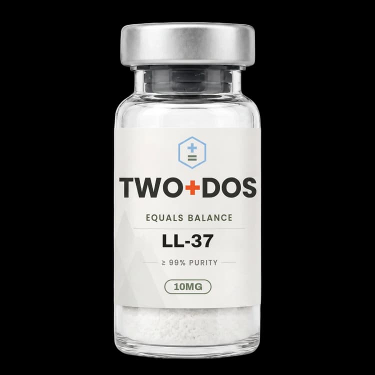
Chemical and physical properties of LL-37 (Cathelicidin)
| Compound class | Cationic α-helical host-defense peptide of the cathelicidin family |
|---|---|
| Residue count | 37 amino acids, corresponding to residues 134-170 of the hCAP18 precursor |
| One-letter sequence | LLGDFFRKSKEKIGKEFKRIVQRIKDFLRNLVPRTES |
| Charged residues | 16 of 37 residues carry charge, giving a net charge of +6 at neutral pH |
| Molecular formula | C205H340N60O53 (PubChem CID 16198951) |
| Average molecular weight | 4493 g/mol |
| Encoding gene | CAMP, human chromosome 3p21.3 — the only cathelicidin gene in the human genome |
| Precursor processing | Released from the 18 kDa hCAP18 pro-protein by proteinase 3 in neutrophils and by kallikreins in keratinocytes |
| Micelle-bound conformation | Curved amphipathic helix-bend-helix spanning residues 2-31, bend between Gly-14 and Glu-16 |
| Smallest active fragment | KR-12, residues 18-29 of the full-length sequence |
| Reported host receptor | Formyl peptide receptor-like 1 (FPRL1, also designated FPR2) |
What is the molecular structure of LL-37?
LL-37 comprises 37 residues with the sequence LLGDFFRKSKEKIGKEFKRIVQRIKDFLRNLVPRTES, a molecular formula of C205H340N60O53 and an average mass near 4493 g/mol under PubChem CID 16198951. Sixteen of those residues carry charge, producing a net charge of +6 at neutral pH and a strongly cationic molecular surface.
Solution structure was resolved by three-dimensional triple-resonance NMR spectroscopy in sodium dodecyl sulfate micelles. Wang reported a curved amphipathic helix-bend-helix motif spanning residues 2-31, with the helical bend located between Gly-14 and Glu-16 and the C-terminal tail remaining disordered. Aromatic residues Phe-5, Phe-6, Phe-17 and Phe-27, together with the arginine side chains, were identified as the contacts engaging anionic lipid headgroups. That geometry is the structural basis of the amphipathicity: cationic and hydrophobic faces separate along the helix axis rather than mixing.
Genetic origin is unusually simple for an antimicrobial peptide family. The CAMP gene at human chromosome 3p21.3 is the single cathelicidin locus in the human genome, and it encodes an 18 kDa pre-protein processed to the 16 kDa hCAP18 propeptide. LL-37 is liberated from the C-terminal domain of that propeptide by extracellular serine protease cleavage, attributed to proteinase 3 in neutrophils and to kallikreins in keratinocytes, so the mature peptide and its inactive reservoir are distinct molecular species in the same compartment.
How does LL-37 interact with bacterial and eukaryotic membranes?
LL-37 associates with membranes through electrostatics before hydrophobic insertion. The +6 net charge drives contact with anionic bacterial surface components, principally lipopolysaccharide in Gram-negative organisms and lipoteichoic acid in Gram-positive organisms, and the amphipathic helix then partitions into the bilayer. The 2025 review by Neshani and colleagues catalogued reported activity spanning more than 38 bacteria, 16 fungi and 16 viruses.
Mechanism at the bilayer is not fixed; it switches with lipid chemistry. Published biophysical work reports two distinct pathways depending on acyl-chain saturation: transmembrane pore formation in bilayers built from unsaturated phospholipids, and membrane modulation in saturated phospholipid systems that instead yields helix-rich fibrous peptide-lipid superstructures. The same peptide therefore produces different lesion geometries in different membrane environments, which complicates any single-mechanism description.
Cholesterol content sets the selectivity margin against host membranes. A 2026 study in Biophysical Chemistry examined LL-37 against eukaryotic-like models built from phosphatidylcholine, sphingomyelin and graded cholesterol fractions using FT-IR, differential scanning calorimetry, isothermal titration calorimetry and fluorescence spectroscopy. LL-37 preferentially perturbed sphingomyelin-enriched membranes, an effect still measurable at 10 mol% cholesterol but markedly diminished at 20 mol%, and the authors described cholesterol as conferring stabilization against peptide-induced disruption.
Bacteria are not passive under this pressure. A 2026 report in Biochimica et Biophysica Acta Biomembranes quantified the lipid response of Staphylococcus aureus exposed to LL-37 and the peptide ATRA-1 using HPLC-MS/MS. Exposure to LL-37 raised membrane rigidity through altered carotenoid content, and both peptides lowered menaquinone and digalactosyl-diacylglycerol levels, with membrane electric surface potential and lipid packing tracked by FTIR. The bilayer that LL-37 encounters after exposure is compositionally different from the one it first met.
What is known about LL-37 oligomerization and aggregation state?
LL-37 does not act as an isolated monomer in most reported systems. Sancho-Vaello and colleagues crystallized the full-length peptide in the presence of detergents and bacterial cell-wall components and resolved a narrow tetrameric channel with a strongly charged core. Cross-linking and molecular dynamics supported tetramer stability, and time-lapse microscopy of Escherichia coli showed membrane discontinuities before cell death.
Aggregation propensity maps onto activity residue by residue. A 2026 Nanoscale study applied discrete molecular dynamics to the LL-37(17-29) fragment and two point mutants. Wild-type peptide assembled into densely packed fibrils held together by hydrophobic helix-helix contacts and retained strong antimicrobial activity; the F17S substitution reduced aggregation and weakened activity; the I24K substitution suppressed aggregation and abolished antimicrobial activity outright, which ties the fibrillar state directly to function rather than treating it as an artifact.
Assembly dynamics also govern selectivity in mixtures. A 2026 report in Angewandte Chemie combined TIRF microscopy, FRET, NMR and molecular dynamics to examine LL-37 together with the human α-defensin HNP1, and described a double cooperativity in which the two peptides raised antimicrobial efficiency while lowering cytotoxicity toward host cells. Lipid composition regulated the aggregation state: anionic lipids dispersed the peptides within the membrane, which the authors linked to selective destruction of bacterial bilayers.
Nucleic acids act as a further switch on assembly. A 2026 eLife study reported that RNA functions as a concentration-dependent regulator of two α-helical peptides with opposite outcomes: it drove the staphylococcal virulence factor PSMα3 through liquid-like condensates into fibrillar polymorphs while cytotoxicity persisted, whereas it attenuated LL-37 cytotoxicity toward host cells while antibacterial function was preserved. The authors framed activity as governed by supramolecular architecture and assembly trajectory rather than by monomer concentration alone.
How does LL-37 engage host receptors and nucleic acids?
LL-37 has a defined host receptor separate from its membrane activity. Yang and colleagues reported in 2000 that the peptide chemoattracts human peripheral blood neutrophils, monocytes and T cells, and induced calcium mobilization in cells transfected with formyl peptide receptor-like 1. That receptor, now commonly designated FPR2, gives LL-37 a signaling role in leukocyte recruitment independent of microbial killing.
Complex formation with DNA is a second, well-documented interaction. Lande and colleagues reported in Nature in 2007 that LL-37 converts inert self-DNA into a trigger of interferon production in plasmacytoid dendritic cells by binding the DNA into aggregated and condensed structures. The peptide is the organizing element in that complex; free DNA in the same system did not elicit the response.
Quantitative description of that condensation arrived in 2026. Zielke and colleagues used phage λ DNA with high-resolution video microscopy, gel electrophoresis, circular dichroism and displacement assays to show non-specific LL-37 binding to double-stranded DNA reaching complete complexation at a DNA to LL-37 weight ratio of 1:1.7. At higher peptide concentrations the complexes adopted disc-like structures roughly 150 nm in diameter, cationic residues engaged the phosphodiester backbone with preference for A-T rich sequences, and the projected area of neutrophil extracellular traps fell measurably at elevated peptide concentrations.
Second-messenger transport is a more recent addition to this picture. A 2025 Cell Reports study described LL-37 carrying immunoreactive cGAMP into cells, activating STING signaling and amplifying interferon-mediated antiviral responses. Read alongside the DNA-condensation work, the pattern is consistent: LL-37 repeatedly acts as a cationic chaperone that delivers anionic cargo across membranes that the cargo could not cross alone.
What have 2025 and 2026 studies reported about LL-37 in host-pathogen systems?
LL-37 appears in recent microbiology literature less as a static killer and more as a molecule that pathogens have evolved specific countermeasures against. Three 2026 reports illustrate the pattern across an enteric bacterium, a Gram-positive pathogen and a non-enveloped virus, each identifying a discrete molecular node rather than a general susceptibility.
Proteolytic evasion was mapped with residue-level precision in a 2026 Science Advances study. Campylobacter jejuni infection induced LL-37 production by intestinal epithelial cells, and the peptide was effective against 86.3% of clinical C. jejuni isolates. Resistant isolates expressed the serine protease HtrA, activated through the NssR transcriptional regulator on exposure to LL-37, which cleaved the peptide at the Ile20-Val21 bond and neutralized its activity. A cleavage-resistant variant, LL-37 I20M/V21R, was constructed and reported to improve bacterial clearance in mouse models.
Interference with bacterial signaling is a separate mode. A 2026 study in Biochemical and Biophysical Research Communications used surface plasmon resonance to show LL-37 binding directly to the CovS sensor kinase of group A Streptococcus, inhibiting its kinase activity in a concentration-dependent manner and antagonizing CovR-mediated activation of the CovRS two-component virulence system. The peptide acts here on a regulatory protein, not on the envelope.
Antiviral work in 2026 identified a post-entry step. A study in Viruses reported that LL-37 reduced Enterovirus 71 replication after entry by activating EGFR-ERK signaling and upregulating the STAC protein; shRNA silencing of STAC increased infection while STAC overexpression decreased it, placing the host protein between peptide exposure and the antiviral outcome.
Host-side measurements from the same window supply context on where the peptide is found. Published quantifications place LL-37 in bronchoalveolar lavage fluid from healthy human donors at roughly 2-5 µg/mL, equivalent to about 0.4-1 µM, rising toward 20 µg/mL during infection, with nasal secretion values reported across a wide 1.2-80 µg/mL range. Laboratory working concentrations in the micromolar range therefore sit near the upper end of what is measured in human fluids.
How do LL-37-derived fragments and analogs compare with the full-length peptide?
LL-37 has been systematically truncated to locate its minimal active core. Wang identified KR-12, spanning residues 18-29, as the smallest fragment retaining antimicrobial activity, folding into a short three-turn amphipathic helix rich in positively charged side chains. In the reported assays KR-12 was bactericidal against Escherichia coli while sparing human cells, establishing the central helix as the functional unit.
Hybrid designs built on that core. A 2026 study in the European Journal of Medicinal Chemistry described KF-22, a fusion of Cathelicidin-BF(1-9) with LL-37(17-29), reporting minimum inhibitory concentrations below 5 µg/mL against both Gram-negative and Gram-positive pathogens, rapid bactericidal kinetics, low resistance induction, and activity against biofilms and persister cells. Mechanistic work attributed the effect to membrane targeting and dissipation of the proton motive force, and mouse plasma biochemistry showed no significant change.
Stereochemical modification is a parallel route. A 2026 Biomolecules study compared two LL-37-derived peptides against Mycobacterium tuberculosis and reported D-LL37 as the more potent, with an IC90 of 18.40 ± 0.39 µM and an IC50 of 10.11 ± 0.60 µM, against 25.44 ± 0.36 µM and 15.45 ± 1.40 µM for the comparator LL37-1. Membrane disruption reached 36-44% at IC90, and RT-qPCR showed upregulation of the P-type ATPase genes ctpF, ctpA and ctpH following exposure.
Comparative biophysics work published in 2025 characterized further LL-37-derived sequences against multidrug-resistant Escherichia coli and ESKAPE organisms, reporting lower minimum inhibitory concentrations and faster bactericidal kinetics than the native peptide alongside low cytotoxicity toward human epithelial and immune cell lines at bactericidal concentrations. Across these reports the design logic is consistent: shorten to the amphipathic core, adjust charge and hydrophobicity, and reduce the proteolytic and ionic-strength liabilities of the full-length sequence.
Summary of published research
Findings below are reported as published by the cited authors, in the model systems they used. They describe laboratory research, and none of them characterize use in humans.
De Yang, Chen Q, Schmidt AP, Anderson GM, Wang JM, Wooters J, Oppenheim JJ, Chertov O. LL-37, the neutrophil granule- and epithelial cell-derived cathelicidin, utilizes formyl peptide receptor-like 1 (FPRL1) as a receptor to chemoattract human peripheral blood neutrophils, monocytes, and T cells. Journal of Experimental Medicine (2000)
- Model system
- primary human leukocytes and FPRL1-transfected kidney cell line
- Conditions
- chemotaxis assays with human peripheral blood neutrophils, monocytes and T cells; calcium mobilization in FPRL1-transfected cells
- Reported finding
- LL-37 was chemotactic for human neutrophils, monocytes and T cells and induced calcium mobilization in monocytes and in kidney cells transfected with formyl peptide receptor-like 1, identifying a defined host receptor through which the peptide recruits leukocytes independently of direct microbial killing.
Lande R, Gregorio J, Facchinetti V, Chatterjee B, Wang YH, Homey B, Cao W, Wang YH, Su B, Nestle FO, Zal T, Mellman I, Schröder JM, Liu YJ, Gilliet M. Plasmacytoid dendritic cells sense self-DNA coupled with antimicrobial peptide. Nature (2007)
- Model system
- human plasmacytoid dendritic cell cultures
- Conditions
- self-DNA presented alone or complexed with LL-37; interferon output as the primary readout
- Reported finding
- LL-37 converted inert self-DNA into a trigger of interferon production by binding the DNA into aggregated and condensed structures, establishing the peptide as the organizing component of an immunostimulatory nucleic-acid complex rather than an inert carrier.
Wang G. Structures of human host defense cathelicidin LL-37 and its smallest antimicrobial peptide KR-12 in lipid micelles. Journal of Biological Chemistry (2008)
- Model system
- cell-free (SDS and dodecylphosphocholine micelles) with bacterial killing assays
- Conditions
- three-dimensional triple-resonance NMR spectroscopy of full-length LL-37 and truncation series down to 12 residues
- Reported finding
- The micelle-bound structure was a curved amphipathic helix-bend-helix spanning residues 2-31 with the bend between Gly-14 and Glu-16 and a disordered C-terminal tail; residues 18-29 (KR-12) were identified as the smallest fragment retaining antibacterial activity, forming a three-turn amphipathic helix that was bactericidal against Escherichia coli while sparing human cells.
Sancho-Vaello E, Gil-Carton D, François P, Bonetti EJ, Kreir M, Pothula KR, Kleinekathöfer U, Zeth K. The structure of the antimicrobial human cathelicidin LL-37 shows oligomerization and channel formation in the presence of membrane mimics. Scientific Reports (2020)
- Model system
- cell-free crystallography, planar lipid bilayers and Escherichia coli imaging
- Conditions
- crystallization with detergents and bacterial cell-wall components; cross-linking, molecular dynamics, conductance measurement and time-lapse microscopy
- Reported finding
- Full-length LL-37 crystallized as a narrow tetrameric channel with a strongly charged core; cross-linking and simulation supported tetramer stability, conductivity was measurable in lipid membranes with water passage observed in simulation, and time-lapse imaging showed membrane discontinuities in Escherichia coli preceding cell death.
Neshani A, Zare H, Ghiasi NS, Karimi MA, Hosseini Bafghi M. Decoding LL-37: Structure and antimicrobial mechanisms against microbial threats. Infection, Genetics and Evolution (2025)
- Model system
- review
- Conditions
- narrative synthesis of structural and antimicrobial mechanism literature across bacterial, fungal and viral systems
- Reported finding
- The review catalogued reported LL-37 activity against more than 38 bacteria, 16 fungi and 16 viruses, and grouped the described mechanisms into cell-wall destruction, membrane permeabilization, oxidative stress, cell-cycle arrest, adhesion blockade, gene expression modulation, and disruption of viral envelopes, entry and replication.
Wei X, Zhang L, Yang Y, Hou Y, Xu Y, Wang Z, et al. LL-37 transports immunoreactive cGAMP to activate STING signaling and enhance interferon-mediated host antiviral immunity. Cell Reports (2025)
- Model system
- cell line and rodent
- Conditions
- cGAMP transfer assays with LL-37; STING pathway activation and interferon output as readouts
- Reported finding
- LL-37 was reported to carry immunoreactive cGAMP into cells, activating STING signaling and amplifying interferon-mediated antiviral responses, extending the peptide's described role as a cationic carrier of anionic cargo across membranes.
Zielke C, Rad B, Nielsen JE, Li J, Pimcharoen S, Sawant M, et al. Human cathelicidin peptide LL-37 compacts nucleic acids and alters neutrophil extracellular trap structure. Scientific Reports (2026)
- Model system
- cell-free (phage λ DNA) and primary human neutrophils
- Conditions
- high-resolution video microscopy, gel electrophoresis, circular dichroism and displacement assays across a range of DNA to peptide weight ratios
- Reported finding
- LL-37 bound double-stranded DNA non-specifically with complete complexation reached at a DNA to LL-37 weight ratio of 1:1.7, forming disc-like structures approximately 150 nm in diameter at higher peptide concentrations; cationic residues engaged the phosphodiester backbone with preference for A-T rich sequences, and projected neutrophil extracellular trap area was reduced at elevated peptide concentrations.
Giraldo-Lorza JM, Hernández Martínez SA, Sierra-Valdez FJ, Leidy C, Suesca E, Manrique-Moreno M. How cholesterol modulates LL-37 function: A biophysical study in eukaryotic-like membrane systems. Biophysical Chemistry (2026)
- Model system
- cell-free (eukaryotic-like model membranes)
- Conditions
- phosphatidylcholine and sphingomyelin membranes with graded cholesterol fractions; FT-IR, differential scanning calorimetry, isothermal titration calorimetry and fluorescence spectroscopy
- Reported finding
- LL-37 preferentially perturbed sphingomyelin-enriched membranes, with the effect still detectable at 10 mol% cholesterol but markedly diminished at 20 mol%, indicating that cholesterol content stabilizes eukaryotic-like bilayers against peptide-induced disruption.
Fuertes-Chaves C, Gonzalez JE, Suesca E, Guzmán-Sastoque P, Muñoz-Camargo C, Manrique-Moreno M. Exposure to the antimicrobial peptides LL-37 and ATRA-1 induces a lipidome response in Staphylococcus aureus that alters membrane biophysical properties. Biochimica et Biophysica Acta Biomembranes (2026)
- Model system
- bacterial culture (Staphylococcus aureus)
- Conditions
- HPLC-MS/MS lipidomics after peptide exposure; membrane electric surface potential and lipid packing assessed by FTIR
- Reported finding
- Exposure to LL-37 increased membrane rigidity through altered carotenoid content, and both LL-37 and ATRA-1 lowered menaquinone and digalactosyl-diacylglycerol levels, demonstrating that the bacterial lipidome remodels in response to peptide challenge rather than remaining a fixed target.
Jin X, Wang H, Tang H. Molecular interaction modes of the host-defense peptide cathelicidin LL-37 and its mutants dictate diverse antimicrobial activities. Nanoscale (2026)
- Model system
- cell-free (discrete molecular dynamics simulation)
- Conditions
- simulation of wild-type LL-37(17-29) alongside the F17S and I24K point mutants
- Reported finding
- Wild-type LL-37(17-29) assembled into densely packed fibrils through hydrophobic helix-helix contacts and retained strong antimicrobial activity, the F17S substitution reduced aggregation and weakened activity, and the I24K substitution suppressed aggregation and abolished antimicrobial activity, linking the fibrillar assembly state directly to function.
Li X, Zhang M, Xu Z, Li Y, Bian S, Tang Y, et al. Serine protease HtrA promotes Campylobacter jejuni intestinal colonization through degrading antimicrobial peptide LL-37. Science Advances (2026)
- Model system
- bacterial clinical isolates, intestinal epithelial cells and mouse colonization model
- Conditions
- susceptibility screening across clinical Campylobacter jejuni isolates; protease cleavage mapping; engineered cleavage-resistant peptide variant
- Reported finding
- LL-37 was effective against 86.3% of clinical Campylobacter jejuni isolates, while resistant isolates expressed the serine protease HtrA, activated through the NssR regulator on peptide exposure, which cleaved LL-37 at the Ile20-Val21 bond; the engineered variant LL-37 I20M/V21R resisted cleavage and improved bacterial clearance in mice.
Santos PA, Maya-Hoyos M, Salazar LM, Cruz CA, Cruz-Cacais A, Giraldo-Avila M, et al. Optimized LL-37-Derived Peptides Exhibit Antitubercular Activity, Induce Membrane Disruption, and P-Type ATPase Transcriptional Responses in Mycobacterium tuberculosis. Biomolecules (2026)
- Model system
- bacterial culture (Mycobacterium tuberculosis)
- Conditions
- growth inhibition assays with D-LL37 and LL37-1; membrane permeabilization measurement at IC90; RT-qPCR of P-type ATPase transcripts
- Reported finding
- D-LL37 returned an IC90 of 18.40 ± 0.39 µM and an IC50 of 10.11 ± 0.60 µM against 25.44 ± 0.36 µM and 15.45 ± 1.40 µM for LL37-1, membrane disruption reached 36-44% at IC90, and exposure upregulated the P-type ATPase genes ctpF, ctpA and ctpH.
What laboratory handling information is published?
LL-37 is supplied as a lyophilized powder, and its physicochemical profile matches that format: an average mass of 4493 g/mol, 16 charged residues out of 37, and a net charge of +6 at neutral pH describe a large, strongly cationic and highly water-soluble solid. Published methods sections prepare aqueous stock solutions in water or dilute buffer before diluting into assay medium.
Assay conditions in the literature are unusually consequential for this molecule. Reported activity is sensitive to ionic strength and to sequestration by serum components, and the mature peptide binds apolipoprotein A-I in human serum while the hCAP18 precursor associates with low-density and very-low-density lipoprotein particles. Comparisons across published values are therefore only meaningful when salt concentration, serum content and peptide-to-lipid or peptide-to-DNA ratio are matched. The 2026 nucleic-acid work is a clear example, where the reported outcome is defined by a DNA to peptide weight ratio of 1:1.7 rather than by peptide concentration alone.
Working concentrations cluster by model type. Antimycobacterial assays reported IC50 and IC90 values between roughly 10 and 26 µM; LL-37-derived analog work reports minimum inhibitory concentrations below 5 µg/mL for optimized sequences; biophysical membrane studies operate at defined peptide-to-lipid ratios in vesicle or monolayer systems with cholesterol fractions specified in mol%. For orientation, quantifications in human bronchoalveolar lavage fluid fall near 2-5 µg/mL, about 0.4-1 µM, in healthy donors.
Analytical confirmation follows standard peptide practice: identity by electrospray mass spectrometry against an average mass of 4493 Da, which for a 37-mer of this charge appears as a multiply protonated envelope rather than a single ion, and purity by reversed-phase HPLC reported on the lot certificate of analysis. Lyophilized material is normally kept frozen, protected from light and moisture. Because highly cationic peptides adsorb to container surfaces, published protocols commonly specify low-binding polypropylene and aliquoting of aqueous stocks so that repeated freeze-thaw cycles are avoided.
This material is offered for laboratory research use only. It is not a drug, not a food, and not intended for human or veterinary use.
Frequently asked research questions
Is LL-37 the same thing as cathelicidin?
LL-37 is the mature, active peptide derived from the human cathelicidin protein, not a separate molecule. The CAMP gene at chromosome 3p21.3 is the only cathelicidin gene in the human genome; it encodes the 18 kDa hCAP18 pre-protein, and LL-37 corresponds to its 37-residue C-terminal domain, residues 134-170, released by extracellular serine protease cleavage.
Why is LL-37 named with two letters and a number?
The designation is descriptive rather than a code. The mature peptide begins with two leucine residues and is 37 amino acids long, giving LL-37. The same convention explains the fragment name KR-12: it starts with lysine and arginine and spans 12 residues, corresponding to positions 18-29 of the parent sequence.
What gives LL-37 selectivity for bacterial over eukaryotic membranes?
Two factors recur in the literature. Bacterial surfaces present anionic lipopolysaccharide and lipoteichoic acid that attract the +6 net charge, while eukaryotic outer leaflets are largely zwitterionic. Cholesterol supplies the second margin: a 2026 biophysical study reported LL-37 perturbation of eukaryotic-like membranes still measurable at 10 mol% cholesterol but markedly diminished at 20 mol%.
Why does aggregation state matter so much in LL-37 research?
Because activity tracks assembly rather than monomer concentration. Crystallography resolved a tetrameric channel, simulation showed that abolishing fibril formation with an I24K substitution abolished antimicrobial activity, and a 2026 eLife report described RNA attenuating host-cell cytotoxicity while antibacterial function persisted. Reported outcomes therefore depend on the supramolecular form present under the specific assay conditions used.
Which model systems dominate the LL-37 literature?
Four recur across the citations above: cell-free lipid vesicle, monolayer and micelle systems for structure and membrane biophysics; bacterial and mycobacterial cultures including clinical isolates for susceptibility and resistance mechanisms; primary human leukocytes and dendritic cells for receptor signaling and nucleic-acid complex work; and rodent colonization and inflammation models for in-vivo readouts.
LL-37 (Cathelicidin) at TWO+DOS
TWO+DOS supplies LL-37 (Cathelicidin) as a research-use-only compound, third-party tested, with certificates of analysis emailed immediately on request.
View the LL-37 (10mg)listing →Related research overviews
References
- PubChem CID 16198951: LL-37 compound summary
- Structures of human host defense cathelicidin LL-37 and its smallest antimicrobial peptide KR-12 in lipid micelles (J Biol Chem 2008)
- The structure of the antimicrobial human cathelicidin LL-37 shows oligomerization and channel formation in the presence of membrane mimics (Sci Rep 2020)
- LL-37, the neutrophil granule- and epithelial cell-derived cathelicidin, utilizes formyl peptide receptor-like 1 (FPRL1) as a receptor (J Exp Med 2000)
- Plasmacytoid dendritic cells sense self-DNA coupled with antimicrobial peptide (Nature 2007)
- Decoding LL-37: Structure and antimicrobial mechanisms against microbial threats (Infect Genet Evol 2025)
- LL-37 transports immunoreactive cGAMP to activate STING signaling and enhance interferon-mediated host antiviral immunity (Cell Rep 2025)
- Human cathelicidin peptide LL-37 compacts nucleic acids and alters neutrophil extracellular trap structure (Sci Rep 2026)
- How cholesterol modulates LL-37 function: A biophysical study in eukaryotic-like membrane systems (Biophys Chem 2026)
- Serine protease HtrA promotes Campylobacter jejuni intestinal colonization through degrading antimicrobial peptide LL-37 (Sci Adv 2026)
- Aggregation-State Dynamics Drive Double Cooperativity Between Antimicrobial Peptides LL-37 and HNP1 (Angew Chem 2026)
- RNA selectively modulates activity of virulent amyloid PSMα3 and host-defense LL-37 via phase separation and aggregation dynamics (eLife 2026)
For research use only. Not for human or veterinary use. Not for diagnostic or therapeutic use.