
VIP (Vasoactive Intestinal Peptide): VPAC1 and VPAC2 Receptor Research
By the TWO+DOS Research Team · Published 2026-08-13
For research use only. Not for human or veterinary use. Not for diagnostic or therapeutic use.
VIP (vasoactive intestinal peptide) is a 28-residue, C-terminally amidated neuropeptide of the secretin/glucagon superfamily, encoded at human chromosome 6q25.2 and excised from a 170-residue precursor. Published research characterises the molecule as the endogenous agonist at the class B1 G protein-coupled receptors VPAC1 and VPAC2, both of which couple to Gs and raise cyclic AMP.
What follows gathers what peer-reviewed sources report about the peptide: verified chemical identifiers and computed physicochemical values, the receptor pharmacology recorded in the IUPHAR literature, the 2022 cryo-EM structure of the receptor-bound peptide, the lymphocyte and enteric-neuron findings that dominate current work, and results from organoid, rodent and cell-line studies published between 2011 and 2026. All material described here is for laboratory research use only.
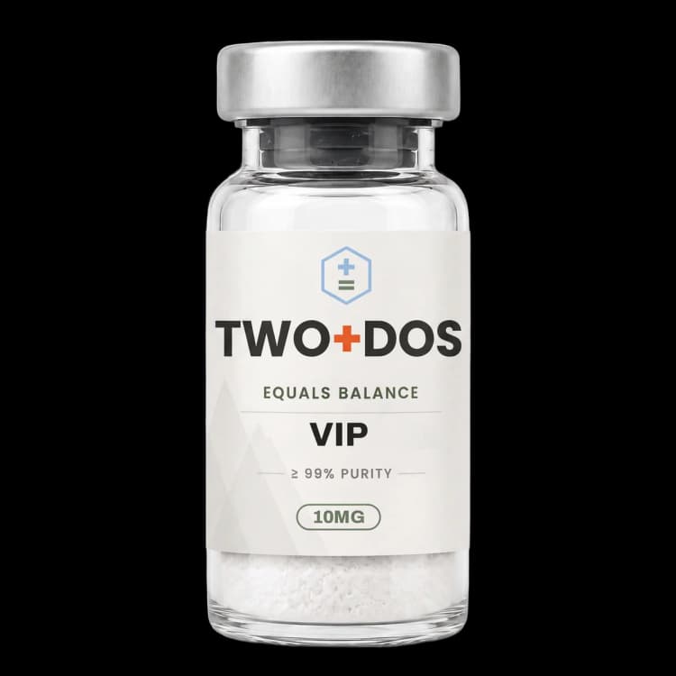
Chemical and physical properties of VIP (vasoactive intestinal peptide)
| Compound class | 28-residue neuropeptide of the secretin/glucagon superfamily; endogenous agonist at class B1 G protein-coupled receptors |
|---|---|
| Residue sequence | His-Ser-Asp-Ala-Val-Phe-Thr-Asp-Asn-Tyr-Thr-Arg-Leu-Arg-Lys-Gln-Met-Ala-Val-Lys-Lys-Tyr-Leu-Asn-Ser-Ile-Leu-Asn, amidated at the C-terminal asparagine |
| Precursor | Excised from a 170-residue prepro-VIP polypeptide at residues 125-152 (UniProt P01282) |
| Gene locus | VIP, human chromosome 6q25.2, NC_000006.12 positions 152,750,796-152,759,759 (NCBI Gene 7432) |
| Cognate receptors | VPAC1 (VIPR1) and VPAC2 (VIPR2), both Gs-coupled; PAC1 binds the peptide far more weakly |
| Reported binding affinity | pKi 8.5-9.8 at VPAC1, 7.8-8.8 at VPAC2 and 6.0-6.3 at PAC1 in cloned human receptor radioligand assays |
| Monoisotopic mass | 3324.7401 Da (PubChem CID 53314964) |
| Computed XLogP | -15.9 (PubChem), describing an extremely hydrophilic polypeptide |
| Topological polar surface area | 1470 Ų (PubChem computed), with 51 hydrogen bond donors and 51 acceptors |
| Reported conformation | Largely unstructured in aqueous buffer; a disordered N-terminal region plus a long α-helix when bound to membrane mimetics |
| Structural coordinates | PDB 8E3Z, the cryo-EM VPAC1-VIP-Gs complex resolved at 2.7 Å |
What is VIP and where does the peptide come from?
VIP was isolated from porcine small intestine in 1970 and has since been located throughout central and peripheral nervous tissue. The mature 28-residue peptide is excised from a 170-residue precursor, prepro-VIP, encoded by the VIP gene at human chromosome 6q25.2, and carries an amide group in place of the free C-terminal carboxylate.
PubChem records the amidated peptide under CID 53314964 with the formula C147H237N43O43S and a molecular weight of 3326.8 g/mol, against CAS registry number 37221-79-7. The single-letter sequence HSDAVFTDNYTRLRKQMAVKKYLNSILN contains one methionine at position 17, two arginines, three lysines and no cysteine, so the molecule has no disulfide bond and no intrinsic constraint on its backbone beyond secondary structure.
The same precursor yields a second bioactive product. UniProt P01282 annotates peptide histidine methionine (PHM-27) at residues 81 to 107 and the mature peptide at residues 125 to 152, both amidated. Because the two are cleaved from one polypeptide, tissue that expresses the gene generally releases both, which is worth noting when interpreting immunoassay data from tissue extracts.
Computed descriptors match the behaviour of a highly polar polypeptide rather than a small molecule. PubChem lists an XLogP of -15.9, a topological polar surface area of 1470 Ų, 51 hydrogen bond donors, 51 hydrogen bond acceptors and 115 rotatable bonds. Nothing in that profile suggests membrane partitioning, and the receptor pharmacology below is correspondingly extracellular.
Which receptors does VIP bind, and with what reported affinity?
VIP acts at two class B1 G protein-coupled receptors, VPAC1 (gene VIPR1) and VPAC2 (gene VIPR2), and far more weakly at the PACAP-preferring receptor PAC1. The IUPHAR review of this receptor family lists radioligand binding affinities at cloned human receptors of pKi 8.5 to 9.8 for VPAC1 and 7.8 to 8.8 for VPAC2.
At PAC1 the same compilation records pKi 6.0 to 6.3, roughly two to three orders of magnitude weaker than at VPAC1. That gap is the basis of the standard pharmacological division of the family: PAC1 is the PACAP-selective receptor, while VPAC1 and VPAC2 accept both endogenous peptides with comparable potency. Any experiment that attributes an effect to one subtype needs subtype-selective tools rather than concentration alone.
Which G proteins each receptor recruits was examined directly in a 2026 BRET study in Jurkat T cells. VPAC1 interacted with both Gαs and Gαq subunits, whereas VPAC2 preferentially engaged Gαs. Both receptors raised cyclic AMP when stimulated, but calcium responses appeared only through VPAC1, which supplies a functional readout that distinguishes the two subtypes in the same cell background.
Selective peptide tools recur across the electrophysiology and immunology literature. PG97-269 is used as a VPAC1 antagonist and PG99-465 as a VPAC2 antagonist, while VIPhyb is the long-standing hybrid antagonist scaffold from which newer, higher-affinity variants have been derived. Reports that use these reagents in parallel are the ones that can assign an observation to a specific receptor subtype.
What does cryo-EM show about how VIP engages VPAC1?
VIP bound to VPAC1 was resolved by cryo-electron microscopy in 2022 at a global resolution of 2.7 Å in complex with heterotrimeric Gs, deposited as Protein Data Bank entry 8E3Z. Companion structures of PACAP27 on VPAC1 and on PAC1 reached 2.3 Å, allowing two related peptides to be compared on the same receptor scaffold.
The comparison is asymmetric in an informative way. The structural analysis counted seven hydrogen bonds between the peptide and VPAC1, against ten for PACAP27 at both receptors, and molecular dynamics simulations showed markedly more extensive movement of the peptide against extracellular loop 3. Fewer stable contacts and a more mobile interface distinguish this ligand from its close relative on the identical receptor.
Solution biophysics had already established why the free peptide is mobile. NMR work published in Biochimica et Biophysica Acta reported that glycine-extended peptide is unstructured in aqueous solution, showing limited chemical shift dispersion, and folds into a disordered N-terminal region followed by a long α-helix only on contact with membrane mimetics. In micelles that helix is curved, with the side chains of Phe6, Tyr10, Leu13 and Met17 forming a hydrophobic patch on the concave face.
Simulations in the 2022 work also modelled the peptide into PAC1 and predicted more transient contacts in that receptor core than in VPAC1, which the authors advance as a structural account of the selectivity gap the binding tables record. Together the structural and solution data explain why substitutions in the C-terminal half of the sequence have been the productive route to higher-affinity antagonists.
How do published studies describe VIP signaling in immune cells?
VIP signaling in lymphocytes is the most heavily worked area of the current literature. Reports describe the peptide raising cyclic AMP through VPAC1 and VPAC2 on T cells, reducing phosphorylation of inflammatory kinase-associated proteins, and acting as an endogenous brake on T cell activation that purpose-built antagonist peptides are designed to lift.
The 2026 Jurkat study built stable lines overexpressing each receptor and read out cyclic AMP, calcium, kinase phosphorylation, immune mediator expression, survival and proliferation. Receptor overexpression alone shifted baseline transcriptional patterns, and peptide exposure lowered phosphorylation of inflammatory kinase-associated proteins in both lines. Proliferation was unchanged, though the VPAC2-overexpressing line showed reduced metabolic activity at 72 hours, and the subtype differences that were clear at early signaling steps converged once the T cell receptor was engaged.
Antagonist chemistry has moved quickly. A 2026 Journal of Biological Chemistry report screened a combinatorial library of C-terminal sequence variants of the VIPhyb scaffold, synthesised 15 candidates, and ranked them by predicted receptor binding, potency in T cell activation assays and activity in murine acute myeloid leukemia models. ANT308 and ANT195 emerged as leads; ANT308 decreased CREB phosphorylation, a downstream readout of receptor engagement, and raised granzyme B and perforin expression in human CD8-positive T cells.
The same axis has been engineered into cell products. A 2026 Science Translational Medicine study reported that the peptide suppresses chimeric antigen receptor T cell function, then built CAR T cells that secrete a short receptor-antagonist peptide. Those cells held a memory phenotype and were metabolically quiescent after manufacture, mounted a strong metabolic response on antigen stimulation, recruited host T cells, and showed greater tumour infiltration in syngeneic and xenogeneic mouse models of haematological and solid tumours.
What do enteric and neuronal VIP studies report?
VIP is expressed by enteric neurons along the length of the gut wall, and 2024 to 2026 work maps a neuroepithelial circuit in which the peptide acts on VIPR1-positive intestinal stem cells. Separate rodent work places peptide-expressing neurons of the suprachiasmatic nucleus at the centre of light-driven changes in memory accessibility.
The Nature Immunology report from 2025 identified peptide-positive enteric neurons acting on VIPR1-positive epithelial stem cells to restrain both proliferation and secretory lineage differentiation. Disrupting that pathway expanded tuft cells, raised interleukin-25 production, activated group 2 innate lymphoid cells and induced a type 2 immune programme resembling worm expulsion. The phenotype was independent of the microbiota but modulated by the IL-25R-ILC2-IL-13 axis and by dietary solids.
Organoid work from 2024 had approached epithelial turnover from the injury side. Jejunal organoids from C57BL/6, Lgr5-EGFP-IRES-CreERT2 and lineage-tracing reporter mice were exposed to the peptide and irradiated at 6 Gy in vitro, with a parallel arm in mice receiving 12 Gy abdominal irradiation. The peptide pushed epithelial differentiation toward secretory phenotypes primarily through p38 MAPK signaling, modulated Lgr5-EGFP-positive progenitor activity, and promoted regeneration after acute irradiation injury.
In the brain, a 2025 Neuroscience Bulletin study reported that acute bright light selectively impaired trace fear memory by activating suprachiasmatic peptide neurons, evidenced by increased c-Fos expression and calcium recording. Optogenetic and chemogenetic manipulation reproduced and reversed the effect, and the projection from the suprachiasmatic nucleus to the paraventricular nucleus of the thalamus proved essential, extending the role of these neurons beyond circadian timing.
Peripheral neuron recordings supply concentration figures. Whole-cell patch-clamp work on myelinated Ah-type trigeminal ganglion neurons from adult female rats reported that 100 nM peptide raised repetitive firing frequency, narrowed the action potential, increased downstroke velocity and deepened after-hyperpolarisation. Iberiotoxin reversed those changes except the after-hyperpolarisation, which apamin abolished, and PG97-269 blocked the response completely while PG99-465 did not, pointing to VPAC1.
What have 2025 and 2026 studies added to the VIP literature?
VIP research published across 2025 and 2026 spans ocular pressure regulation, viral entry biology, lymphocyte signaling and peripheral neuron excitability. Working concentrations differ by orders of magnitude between those systems, from 100 nM in single-neuron electrophysiology upward, so a figure quoted without its model system carries very little information.
Ocular work published in Annals of Medicine in 2025 used two paired systems: rats in which three episcleral veins were cauterised, and human trabecular meshwork cells under pressure stimulation. Both models raised the tight junction proteins ZO-1 and Claudin-1. Peptide exposure and Rab13 overexpression each lowered those proteins in pressure-stimulated cells, Rab13 knockdown reversed the peptide effect, and in the rat model the peptide reduced ZO-1, Claudin-1 and trabecular tissue density alongside lower intraocular pressure.
A 2025 International Journal of Molecular Sciences study examined the peptide against SARS-CoV-2 entry machinery in CaCo-2 epithelial cells stimulated with spike protein. The peptide downregulated ACE2 and TMPRSS2 at both the messenger RNA and surface protein level, and separately drove shedding of the surface-expressed proteins by upregulating the sheddase ADAM10. Infection rate by a SARS-CoV-2 pseudovirus fell as a result, and the authors flag the ADAM10 link as the generalisable mechanism.
Read together, the recent record is notable for how consistently receptor subtype identity, rather than peptide concentration, decides the reported outcome. The 2026 lymphocyte work separates VPAC1 from VPAC2 by calcium coupling, the trigeminal recordings separate them by antagonist reversal, and the antagonist and CAR T reports make receptor engagement itself the variable under study. That convergence is the most useful organising fact for anyone reading the field as of August 2026.
Summary of published research
Findings below are reported as published by the cited authors, in the model systems they used. They describe laboratory research, and none of them characterize use in humans.
Harmar AJ, Fahrenkrug J, Gozes I, Laburthe M, May V, Pisegna JR, Vaudry D, Vaudry H, Waschek JA, Said SI. Pharmacology and functions of receptors for vasoactive intestinal peptide and pituitary adenylate cyclase-activating polypeptide: IUPHAR review 1. British Journal of Pharmacology (2012)
- Model system
- review of cloned human receptor pharmacology
- Conditions
- compilation of radioligand binding affinities at cloned human VPAC1, VPAC2 and PAC1 receptors
- Reported finding
- The review tabulated affinities of pKi 8.5 to 9.8 at VPAC1, 7.8 to 8.8 at VPAC2 and 6.0 to 6.3 at PAC1, establishing the two- to three-order-of-magnitude selectivity gap between the VPAC subtypes and the PACAP-preferring receptor, and noted that the precursor polypeptide encodes several additional bioactive peptides beyond the mature 28-residue chain.
Umetsu Y, Tenno T, Goda N, Shirakawa M, Ikegami T, Hiroaki H. Structural difference of vasoactive intestinal peptide in two distinct membrane-mimicking environments. Biochimica et Biophysica Acta (2011)
- Model system
- cell-free solution NMR
- Conditions
- glycine-extended peptide in two distinct membrane-mimicking environments, compared against aqueous solution
- Reported finding
- The peptide was unstructured in aqueous solution as judged by limited chemical shift dispersion, and adopted a disordered N-terminal region with a long α-helix in both membrane mimetics; the micelle-bound helix was curved, with Phe6, Tyr10, Leu13 and Met17 forming a hydrophobic patch on the concave face oriented toward the micelle interior, the same residue set that forms the micelle-binding interface in PACAP-38.
Piper SJ, Deganutti G, Lu J, Zhao P, Liang YL, Lu Y, Fletcher MM, Hossain MA, Christopoulos A, Reynolds CA, Danev R, Sexton PM, Wootten D. Understanding VPAC receptor family peptide binding and selectivity. Nature Communications (2022)
- Model system
- cryo-electron microscopy plus molecular dynamics simulation
- Conditions
- active, Gs-coupled VIP-VPAC1R, PACAP27-VPAC1R and PACAP27-PAC1R complexes; global map resolutions of 2.7 Å, 2.3 Å and 2.3 Å respectively
- Reported finding
- Structural analysis counted seven hydrogen bonds between the peptide and VPAC1R against ten for PACAP27 at both receptors, and simulations showed more extensive dynamics of the peptide against extracellular loop 3; modelling the peptide into PAC1R predicted more transient contacts in the receptor core than in VPAC1R, offering a structural basis for the observed subtype selectivity. Coordinates were released as PDB 8E3Z.
Agibalova T, Hempel A, Maurer HC, Ragab M, Ermolova A, Wieland J, Waldherr Ávila de Melo C, Heindl F, Giller M, Fischer JC, Tschurtschenthaler M, Kohnke-Ertel B, Öllinger R, Steiger K, Demir IE, Saur D, Quante M, Schmid RM, Middelhoff M. Vasoactive intestinal peptide promotes secretory differentiation and mitigates radiation-induced intestinal injury. Stem Cell Research and Therapy (2024)
- Model system
- murine jejunal organoids and rodent irradiation model
- Conditions
- organoids from C57BL/6, Lgr5-EGFP-IRES-CreERT2 and lineage-tracing reporter mice; 6 Gy irradiation in vitro and 12 Gy abdominal irradiation in vivo
- Reported finding
- The peptide promoted epithelial differentiation toward secretory phenotypes primarily through p38 MAPK signaling and modulated proliferation and Lgr5-EGFP-positive progenitor activity; after acute irradiation injury it promoted epithelial regeneration in organoids, with the effect confirmed in the abdominal irradiation model.
Su X, Tang Y, Zhong Y, Liu Y. Suprachiasmatic Nucleus Vasoactive Intestinal Peptide Neurons Mediate Light-induced Transient Forgetting. Neuroscience Bulletin (2025)
- Model system
- rodent (mouse suprachiasmatic nucleus circuit mapping)
- Conditions
- acute bright light exposure with c-Fos mapping, calcium recording, and optogenetic and chemogenetic manipulation of peptide-expressing suprachiasmatic neurons
- Reported finding
- Acute bright light selectively impaired trace fear memory by activating peptide-expressing suprachiasmatic neurons, shown by raised c-Fos expression and calcium recording; the effect was reproduced and reversed by optogenetic and chemogenetic manipulation, and the suprachiasmatic-to-paraventricular thalamic projection was required, identifying a role in memory accessibility distinct from circadian timing.
Chen L, Yan X, Luo Z, Cheng Y, Li M. Vasoactive intestinal peptide reduces ocular hypertension by regulating tight junction of trabecular meshwork through Rab13/PKA signalling complex. Annals of Medicine (2025)
- Model system
- rodent ocular hypertension model plus human trabecular meshwork cells
- Conditions
- cautery of three episcleral veins in rats and pressure stimulation of human trabecular meshwork cells; ZO-1, Claudin-1, Rab13 and PKA measured by Western blot, qRT-PCR, immunofluorescence and immunohistochemistry
- Reported finding
- Both models raised the tight junction proteins ZO-1 and Claudin-1; the peptide and Rab13 overexpression each lowered them in pressure-stimulated cells while Rab13 knockdown reversed the peptide effect, and in the rat model the peptide reduced ZO-1, Claudin-1 and trabecular tissue density with a corresponding fall in intraocular pressure.
Gutzler C, Höhne K, Bani D, Kayser G, Fähndrich S, Ambros M, Hug MJ, Rieg S, Falcone V, Müller-Quernheim J, Zissel G, Frye BC. Vasoactive Intestinal Peptide (VIP) in COVID-19 Therapy-Shedding of ACE2 and TMPRSS2 via ADAM10. International Journal of Molecular Sciences (2025)
- Model system
- human cell line (CaCo-2 epithelial cells)
- Conditions
- cells stimulated with SARS-CoV-2 spike protein and exposed to the native peptide; ACE2 and TMPRSS2 messenger RNA and surface expression, TMPRSS2 enzyme activity and pseudovirus infection rate measured
- Reported finding
- The peptide downregulated ACE2 and TMPRSS2 at both messenger RNA and surface level and additionally drove shedding of the surface-expressed proteins through upregulation of the sheddase ADAM10; the combined effect lowered infection rate by a SARS-CoV-2 pseudovirus in the same system.
Jakob MO, Sterczyk N, Boulekou S, Forster PM, Barleben L, Alzain N, Jarick KJ, Pirzgalska RM, Diefenbach A, Klose CSN, et al. Enteric nervous system-derived VIP restrains differentiation of LGR5+ stem cells toward the secretory lineage impeding type 2 immune programs. Nature Immunology (2025)
- Model system
- rodent (mouse intestinal neuroepithelial circuit)
- Conditions
- genetic disruption of the peptide-VIPR1 axis in LGR5-positive epithelial stem cells, with microbiota-depleted and diet-modified comparison arms
- Reported finding
- Peptide-positive enteric neurons acted on VIPR1-positive epithelial stem cells to restrain proliferation and secretory lineage differentiation; disrupting the pathway expanded tuft cells, raised interleukin-25, activated group 2 innate lymphoid cells and induced a type 2 immune programme resembling worm expulsion, independent of the microbiota but modulated by the IL-25R-ILC2-IL-13 axis and by dietary solids.
Cabrera-Martín A, Arribas-Castaño P, Castro-Vázquez D, Tecza K, Pérez-García S, Martínez C, El Khamlichi C, Morisset-Lopez S, Juarranz Y, Gutiérrez-Cañas I, Villanueva-Romero R. Finding functional gaps: integrative analysis of VPAC1- and VPAC2-mediated signalling pathways in human lymphocytes. Cell and Bioscience (2026)
- Model system
- human cell line (Jurkat T cells stably overexpressing VPAC1 or VPAC2)
- Conditions
- BRET assays for receptor-G protein interaction; cyclic AMP, calcium, kinase phosphorylation, immune mediator expression, survival and proliferation read across 72 hours
- Reported finding
- VPAC1 interacted with both Gαs and Gαq while VPAC2 preferentially engaged Gαs; both receptors raised cyclic AMP but calcium responses appeared only through VPAC1, peptide exposure lowered phosphorylation of inflammatory kinase-associated proteins in both lines, proliferation was unchanged, the VPAC2 line showed reduced metabolic activity at 72 hours, and subtype differences that were clear at early signaling steps converged after T cell receptor activation.
Lin HK, Blake DA, Freeman R, Chen Y, Kim J, Johnson AM, Wells K, Mudigonda A, Waller EK, Rafiq S, et al. Modulating the VIP-VIPR pathway reprograms CAR T cells for superior antitumor efficacy in preclinical cancer models. Science Translational Medicine (2026)
- Model system
- engineered human CAR T cells plus syngeneic and xenogeneic mouse tumour models
- Conditions
- CAR T cells engineered to secrete a short receptor-antagonist peptide (CAR/VIPRa), compared against unmodified CAR T cells in haematological and solid tumour models
- Reported finding
- The peptide suppressed CAR T cell function, and antagonist-secreting CAR T cells retained a memory phenotype and metabolic quiescence after manufacture while mounting a strong metabolic response to antigen; they recruited host T cells, showed greater tumour infiltration, remained less exhausted and produced better tumour control in both syngeneic and xenogeneic models.
Wang Y, Sen-Majumdar A, Li JM, Sarkar S, Passang T, Mecorapaj S, Balaji S, Kalsang T, Ward AB, Li Y, Cohen J, Chen Z, Chaudagar K, Das PK, Wang S, Bruk N, Papadantonakis N, Giver CR, Waller EK. Identification and characterization of vasoactive intestinal peptide receptor antagonists with high-affinity and potent anti-leukemia activity. Journal of Biological Chemistry (2026)
- Model system
- in silico docking, purified mouse and human T cells, and murine acute myeloid leukemia models
- Conditions
- combinatorial library of VIPhyb C-terminal sequence variants screened by docking against human VPAC1 and VPAC2; 15 peptides synthesised and ranked by T cell activation EC50 and activity in C57BL/6 mice engrafted with the C1498 leukemia line
- Reported finding
- Predicted receptor binding correlated positively with the ability of each variant to augment mouse T cell proliferation and anti-leukemia activity; ANT308 and ANT195 emerged as leads on combined docking score, low in vitro T cell activation EC50 and in vivo activity, and ANT308 decreased CREB phosphorylation, a downstream receptor readout, while raising granzyme B and perforin expression in human CD8-positive T cells.
Yan F, Zhong-Xuan P, Li-Li T, Luo F, Hai-Ying D. Effects of vasoactive intestinal peptide on neuroexcitability of female-specific subpopulation of myelinated Ah-type neurons isolated from trigeminal ganglia of adult female rats. Neuropeptides (2026)
- Model system
- rodent primary neurons (myelinated Ah-type trigeminal ganglion neurons, adult female rats)
- Conditions
- whole-cell patch-clamp recording of action potentials prior to and following exposure to 100 nM peptide, with iberiotoxin, apamin, PG97-269 (VPAC1 antagonist) or PG99-465 (VPAC2 antagonist)
- Reported finding
- 100 nM peptide significantly raised repetitive firing frequency with narrowed action potentials, increased downstroke derivative velocity and deeper after-hyperpolarisation; iberiotoxin reversed all of those changes except the after-hyperpolarisation, which apamin abolished, and PG97-269 completely blocked the firing and repolarisation effects while PG99-465 did not, implicating VPAC1.
What laboratory handling information is published?
VIP is supplied as a lyophilized powder, and the computed profile matches that format precisely. An XLogP of -15.9, a topological polar surface area of 1470 Ų and 51 hydrogen bond donors describe an extremely hydrophilic solid with no lipophilic character, so published methods prepare working solutions in aqueous buffer, saline or culture medium rather than organic solvent.
Peptide solutions of this class adsorb to glass and untreated plastic at low concentrations, which is why cell-assay protocols commonly include carrier protein in the diluent and why the peptide is normally aliquoted rather than repeatedly thawed. The molecule has no cysteine and therefore no disulfide to scramble, but the single methionine at position 17 is an oxidation-sensitive residue and sits inside the hydrophobic patch identified in the NMR work, so oxidative conditions are the chemistry most likely to matter.
Circulating stability is the practical constraint the literature works around. Reviews of this peptide family describe rapid clearance and enzymatic breakdown as the reason so much of the delivery work uses liposomal encapsulation, nanoparticle carriers or sequence-modified analogues; the 2026 antagonist screen explicitly listed plasma stability alongside receptor affinity as a design target for its C-terminal variants.
Working concentrations in the published record vary widely by model type, so a figure quoted without its system is not interpretable. Trigeminal neuron electrophysiology used 100 nM, and the receptor binding tables put affinities in the low nanomolar range, consistent with pKi 8.5 to 9.8 at VPAC1; cell-culture and organoid protocols generally sit well above those figures because the peptide is consumed and degraded over a multi-day incubation. Identity is confirmed by electrospray mass spectrometry against a monoisotopic mass of 3324.7401 Da, which for a 3.3 kDa peptide appears as a series of multiply charged ions, with purity determined by reversed-phase HPLC and reported on the lot certificate of analysis.
This material is offered for laboratory research use only. It is not a drug, not a food, and not intended for human or veterinary use.
Frequently asked research questions
What does the abbreviation VIP stand for?
VIP stands for vasoactive intestinal peptide, also written vasoactive intestinal polypeptide. The molecule is recorded under CAS registry number 37221-79-7 and PubChem CID 53314964, with the sequence HSDAVFTDNYTRLRKQMAVKKYLNSILN and an amide group at the C-terminal asparagine. Older literature also lists it as the synthetic porcine peptide, since the porcine and human sequences are identical.
Which receptors does VIP activate?
Two class B1 G protein-coupled receptors carry most of the pharmacology: VPAC1, encoded by VIPR1, and VPAC2, encoded by VIPR2. Both couple to Gs and raise cyclic AMP. The IUPHAR compilation lists pKi 8.5 to 9.8 at VPAC1 and 7.8 to 8.8 at VPAC2, against 6.0 to 6.3 at the PACAP-preferring PAC1 receptor.
Is VIP the same molecule as PACAP?
No. Pituitary adenylate cyclase-activating polypeptide is a separate member of the same secretin/glucagon superfamily. The two share the VPAC1 and VPAC2 receptors with comparable affinity, but PACAP binds PAC1 two to three orders of magnitude more tightly. Cryo-EM structures of both peptides on VPAC1 showed seven hydrogen bonds for one and ten for the other, so their binding modes differ even on a shared receptor.
How large is the VIP molecule and what shape does it take?
The peptide is 28 residues with a molecular weight of 3326.8 g/mol and the formula C147H237N43O43S. NMR work reported that it is largely unstructured in aqueous solution and folds into a disordered N-terminal region followed by a long α-helix on contact with membrane mimetics, with Phe6, Tyr10, Leu13 and Met17 forming a hydrophobic patch on the concave face of the curved helix.
Which model systems dominate the VIP literature?
Four recur across the citations above: human and murine T cell systems including Jurkat lines and engineered CAR T cells, murine intestinal organoids and enteric neuroepithelial circuits, rodent neuron recordings and brain circuit mapping, and epithelial cell lines such as CaCo-2 and human trabecular meshwork cells. Cryo-EM and solution NMR supply the cell-free structural record.
VIP (vasoactive intestinal peptide) at TWO+DOS
TWO+DOS supplies VIP (vasoactive intestinal peptide) as a research-use-only compound, third-party tested, with certificates of analysis emailed immediately on request.
View the VIP (10mg)listing →Related research overviews
References
- PubChem CID 53314964: vasoactive intestinal peptide compound summary
- UniProt P01282: VIP peptides precursor (Homo sapiens), 170 residues
- NCBI Gene 7432: VIP vasoactive intestinal peptide, chromosome 6q25.2
- RCSB PDB 8E3Z: cryo-EM structure of the VPAC1R-VIP-Gs complex at 2.7 Å
- Pharmacology and functions of receptors for vasoactive intestinal peptide and pituitary adenylate cyclase-activating polypeptide: IUPHAR review 1 (Br J Pharmacol 2012)
- Understanding VPAC receptor family peptide binding and selectivity (Nat Commun 2022)
- Structural difference of vasoactive intestinal peptide in two distinct membrane-mimicking environments (Biochim Biophys Acta 2011)
- Vasoactive intestinal peptide promotes secretory differentiation and mitigates radiation-induced intestinal injury (Stem Cell Res Ther 2024)
- Enteric nervous system-derived VIP restrains differentiation of LGR5+ stem cells toward the secretory lineage impeding type 2 immune programs (Nat Immunol 2025)
- Vasoactive intestinal peptide reduces ocular hypertension by regulating tight junction of trabecular meshwork through Rab13/PKA signalling complex (Ann Med 2025)
- Identification and characterization of vasoactive intestinal peptide receptor antagonists with high-affinity and potent anti-leukemia activity (J Biol Chem 2026)
- Modulating the VIP-VIPR pathway reprograms CAR T cells for superior antitumor efficacy in preclinical cancer models (Sci Transl Med 2026)
For research use only. Not for human or veterinary use. Not for diagnostic or therapeutic use.