
Sermorelin: GRF(1-29) Chemistry, Receptor Data and Peptide Stability
By the TWO+DOS Research Team · Published 2026-08-13
For research use only. Not for human or veterinary use. Not for diagnostic or therapeutic use.
Sermorelin is a synthetic 29-residue peptide reproducing the N-terminal fragment of human growth hormone-releasing hormone, with the formula C149H246N44O42S and a molecular weight of 3357.9 g/mol. It carries a C-terminal amide and acts as an agonist at the GHRH receptor, a class B1 G-protein-coupled receptor found on anterior pituitary somatotrophs.
Two bodies of work define what is measurable about this molecule. The first is peptide chemistry from the late 1980s and early 1990s, which mapped exactly where the sequence falls apart, enzymatically and chemically, with rate constants attached. The second is contemporary analytical chemistry, where the fragment and its N-terminally clipped metabolite are routine targets in anti-doping method development through 2026. Structural biology of the receptor sits between them, most recently in a 2026 letter reporting ligand-free and small-molecule-bound receptor complexes.
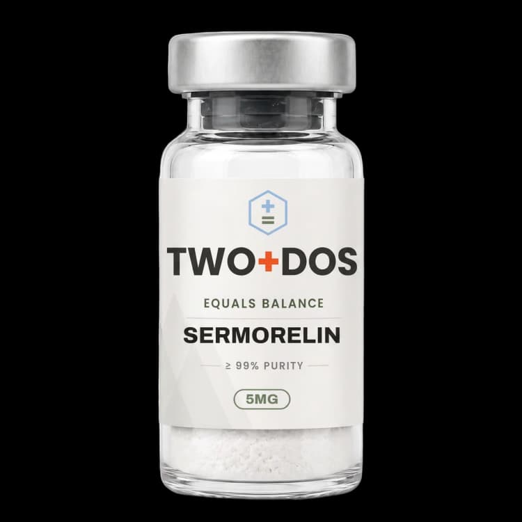
Chemical and physical properties of Sermorelin
| Peptide class | Unmodified 29-residue N-terminal fragment of human growth hormone-releasing hormone, C-terminally amidated; agonist at the GHRH receptor |
|---|---|
| Sequence | YADAIFTNSYRKVLGQLSARKLLQDIMSR-NH2 (29 residues, all L-configuration, primary amide at Arg29) |
| Molecular formula | C149H246N44O42S (free peptide). The acetate salt is listed as C149H246N44O42S · C2H4O2 at 3417.97 g/mol |
| Molecular weight | 3357.9 g/mol; exact mass 3356.8221 Da; monoisotopic mass 3355.8187 Da (PubChem computed) |
| CAS numbers | 86168-78-7 (free peptide); 114466-38-5 is the number circulated for the acetate salt |
| PubChem CID | 16132413 |
| InChIKey | WGWPRVFKDLAUQJ-MITYVQBRSA-N |
| Registry identifiers | UNII 89243S03TE; ChEBI 9118; DTXSID70903978; ATC codes H01AC04 and V04CD03 |
| Computed descriptors | XLogP -12.1; topological polar surface area 1470 square angstroms; 52 hydrogen-bond donors; 49 acceptors; 118 rotatable bonds; 236 heavy atoms; structural complexity 7640 (PubChem computed) |
| Molecular target | GHRH receptor (GHRHR), a 423-residue class B1 G-protein-coupled receptor of the secretin family, coupled to Gs and adenylate cyclase |
| Principal metabolite | Sermorelin(3-29)-NH2, generated by dipeptidyl peptidase IV cleavage of the Ala2-Asp3 bond; carried as a separate analytical target in anti-doping methods |
What is sermorelin?
Sermorelin is the C-terminally amidated 29-residue N-terminal fragment of human growth hormone-releasing hormone, catalogued as GRF(1-29)NH2, hGHRH(1-29)NH2 and Groliberin. Every residue is a standard L-amino acid in the native order, so the molecule is a truncation of the endogenous 44-residue hormone rather than an engineered analogue, and its formula is C149H246N44O42S at 3357.9 g/mol.
The residue numbering matters more here than in most peptides, because four specific positions carry the whole reactivity profile. Ala2 defines the dipeptidyl peptidase IV cleavage site. Asp3 is the residue that isomerises in aqueous solution. Asn8 is the deamidation site. Met27 is the only sulfur atom in the molecule and therefore the only oxidation-sensitive side chain. Lys12 and Lys21 are the two free primary amines that conjugation chemistry targets. Each of those assignments comes from published work summarised in the sections below.
Because the sequence is native, sermorelin has no D-amino acids, no non-proteinogenic residues and no appended chemistry. That makes it the reference point against which the modified members of this peptide family are described, and it is why the chemical literature frequently reports sermorelin numbers alongside those for longer or substituted relatives such as GRF(1-44)-NH2 and GRF(1-20)-NH2.
The unambiguous identifiers are PubChem CID 16132413, InChIKey WGWPRVFKDLAUQJ-MITYVQBRSA-N and CAS 86168-78-7 for the free peptide. Records that describe the acetate carry the composite formula C149H246N44O42S · C2H4O2 at 3417.97 g/mol, roughly 60 g/mol heavier. Documentation should state which of the two a given container holds, since peptide content per milligram differs between them.
How does sermorelin engage the GHRH receptor?
Sermorelin retains the complete recognition determinant of full-length GHRH, so it engages the GHRH receptor through the same interface as the endogenous hormone. Gaylinn and colleagues cloned that receptor from a human pituitary library in 1993: a 423-amino-acid seven-transmembrane protein of the secretin family, sharing 47 percent identity with the vasoactive intestinal peptide receptor.
Class B1 peptide recognition proceeds in two stages: the C-terminal half of the ligand docks against the receptor extracellular domain, then the N-terminal residues descend into the transmembrane bundle and trigger activation. The 29-residue fragment carries both halves of that pharmacophore. Sequence identity with the remaining members of the family falls off from there, at 42 percent for the secretin receptor, 35 percent for calcitonin and 28 percent for parathyroid hormone. Zhou and colleagues resolved the arrangement in 2020 by cryo-electron microscopy of the human receptor with GHRH and heterotrimeric Gs at 2.6 angstroms, showing the helical peptide contacting the extracellular domain, every extracellular loop, and all transmembrane helices except helix 4.
A 2026 letter in Protein and Cell added the states that the 2020 work did not cover. Wang and colleagues determined a ligand-free receptor complex at 3.04 angstroms and a complex with the small-molecule agonist PCO371 at 2.88 angstroms, and set both against the peptide-bound form and against splice variant 1 of the receptor. Those two structures define the conformational baseline and an allosteric entry point distinct from the orthosteric peptide site the 29-residue fragment occupies.
Downstream coupling is to Gs and adenylate cyclase, and cyclic AMP accumulation is the standard functional readout in receptor-expressing cell lines. No published structure shows sermorelin itself bound to the receptor. The structural inferences above are drawn from complexes with the full-length hormone whose first 29 residues sermorelin reproduces exactly, which is a strong basis but still an inference rather than a direct measurement.
Why is the sermorelin sequence short-lived in plasma?
Sermorelin carries an alanine at position 2, and that single residue is the reason the peptide disappears from plasma within minutes. Dipeptidyl peptidase IV removes the N-terminal Tyr-Ala dipeptide, converting the molecule into sermorelin(3-29)-NH2, a fragment that has lost the N-terminal residues the receptor transmembrane bundle recognises.
Bongers and colleagues quantified the reaction in 1992 using purified human placental enzyme with HPLC readout. Working at 37 degrees Celsius and pH 7.8 from a starting substrate concentration of 0.15 mM, they recorded initial cleavage rates near 5 micromoles per minute per milligram of enzyme, and found the same figure for GRF(1-44)-NH2, GRF(1-29)-NH2 and GRF(1-20)-NH2. Truncating the chain from 44 residues to 20 therefore does nothing to protect the scissile bond.
The same study mapped what the enzyme will and will not accept. Analogues bearing a D-configuration residue at P2, P1 or P1', along with desamino-Tyr1 and N-methyl-Tyr1 variants, resisted cleavage entirely. Across a substituted series, catalytic efficiency peaked for a hydrophobic side chain at position 2 measuring roughly 0.25 nanometres, matching alpha-aminobutyric acid. Related work on bovine sequences put relative cleavage rates at 100 percent for Ala2, 4 percent for Ser2, 2.5 percent for Thr2, 0.53 percent for Val2 and zero for Ile2, which is the quantitative basis for every position-2 substitution in this peptide family.
Cleavage products of this family are analytically important even where they are pharmacologically inert. Memdouh and colleagues built sermorelin(3-29)-NH2 into their reference panel precisely because the metabolite persists in a matrix where the parent may already be gone, and later urine methods carry it as a named target alongside the intact peptide.
Which chemical degradation routes has the sequence shown in solution?
Sermorelin degrades in aqueous solution by three routes independent of any enzyme: isomerisation at Asp3, deamidation at Asn8, and oxidation at Met27. Bongers and colleagues isolated the products of the first two from GRF(1-29)-NH2 in 1992 and reported that the peptide is most stable between pH 4 and pH 5.
The pH dependence has a clean chemical explanation, and it held across solutions from 15 to 200 micromolar. Below pH 4 the Asp3 side chain is protonated and the dominant route is chain cleavage at the Asp3-Ala4 bond, yielding GRF(4-29)-NH2. Above pH 4 the ionised carboxylate instead attacks the backbone, and isomerisation to beta-Asp3 predominates. Both processes follow simple first-order kinetics whose rates track pH and are independent of buffer concentration.
Asn8 deamidation is the faster of the two and proceeds through a cyclic imide intermediate at pH 5 and above, generating beta-Asp8 and Asp8 products in roughly a 4:1 ratio under general base catalysis. Below pH 2 a separate direct hydrolysis of the protonated amide gives the alpha-aspartyl product exclusively. Notably, deamidation at pH 8.0 proved relatively insensitive to temperature below 37 degrees Celsius, which the authors attributed to conformational constraint. Asp25 and Asn35 showed no comparable degradation under the same conditions.
The potency cost of deamidation was measured on a closely related analogue. Friedman and colleagues reported in 1991 that the beta-Asp8 product of a GRF(1-32)NH2 analogue was 400 to 500 times weaker than the parent in cultured bovine anterior pituitary cells, and the alpha-Asp8 product 25 times weaker. In phosphate buffer at pH 7.4 and 37 degrees Celsius the parent peptide showed a disappearance half-life of 202 hours, while replacing Asn8 with serine extended it to 1550 hours. Oxidation of Met27 to the sulfoxide is the third documented route and is the reason substituted analogues in this family commonly carry leucine at that position.
What pharmacokinetic values have been reported in human volunteers?
Sermorelin has published human clearance figures from constant-infusion work. Soule and colleagues infused GHRH-(1-29)-NH2 into ten normal men at 25 nanograms per kilogram per minute and reported a metabolic clearance rate of 39.7 plus or minus 3.9 millilitres per kilogram per minute and a half-life of 4.3 plus or minus 1.4 minutes.
The comparison built into that study is what makes the numbers interpretable. A D-Ala2 analogue run under identical conditions cleared at 21 plus or minus 1.2 millilitres per kilogram per minute, significantly slower at P below 0.001, with a half-life of 6.7 plus or minus 0.5 minutes. Inverting one stereocentre at the dipeptidyl peptidase IV site therefore cut clearance roughly in half, which links the plasma figure directly back to the enzymology.
Other reported values sit in the same region. A 2003 review of GRF conjugation chemistry by Esposito and colleagues described the plasma half-life of GRF(1-29) in humans as approximately 10 to 20 minutes, a range consistent with the infusion data once differences in sampling and assay are allowed for. That review also identified Lys12 and Lys21 as the positions where mono-PEGylated conjugates carrying a 5000-dalton polymer retained in vitro activity comparable to the unmodified peptide.
On the pharmacodynamic side, Wilton and colleagues studied thirty healthy men aged 19 to 43 and found that 0.25 micrograms per kilogram was already sufficient to produce a measurable rise in circulating growth hormone, with peaks near 90 milliunits per litre at 1 to 2 micrograms per kilogram. The same study measured intranasal bioavailability at only 3 to 5 percent, an unsurprising figure for a 3.4 kDa peptide with a computed XLogP of minus 12.1 and 52 hydrogen-bond donors.
How is sermorelin detected and confirmed analytically?
Sermorelin is confirmed by liquid chromatography coupled to tandem or high-resolution mass spectrometry, verified against a monoisotopic mass of 3355.8187 Da and an average molecular weight of 3357.9 g/mol. Anti-doping laboratories have driven most method development for this compound, and that literature is where the quantitative sensitivity figures live.
Four method papers bracket the current position. Memdouh and colleagues incubated four of the larger GHRH-family analogues in fortified urine in 2021, characterised nineteen major in vitro metabolites, synthesised them as reference materials, and reached detection limits generally at or below 1 nanogram per millilitre. Cristea and colleagues published a weak cation-exchange extraction with triple-quadrupole instrumentation in 2023, reporting a detection limit of 0.2 and a quantification limit of 0.6 nanograms per millilitre with linearity from 0.1 to 1.2. Thomas and colleagues consolidated the entire 2 to 10 kilodalton analyte window into one extraction in 2024, controlled by five internal standards. Ucakturk and Nemutlu combined ultrafiltration with solid-phase extraction ahead of nano-flow chromatography and Orbitrap detection in 2026, reaching detection limits of 0.5 nanograms per millilitre or better and identification limits between 0.5 and 0.75.
Fragment behaviour under proteolysis has been characterised separately, and one result is directly useful in the laboratory. Gonzalez-Lopez and colleagues synthesised a panel of doping-relevant peptides by solid-phase chemistry in 2023 and challenged them with human blood, trypsin and chymotrypsin under HPLC-DAD monitoring. The C-terminal octapeptide sermorelin(22-29) survived all three challenges intact, which qualified it as an in-house internal standard for quantification work in biological matrices.
Two constraints recur across all of these methods. Urinary concentrations of this peptide family are small, which is why preconcentration and nano-flow separations keep appearing. And the analyte is fragile in matrix, so extraction chemistry and cold handling carry as much weight in the reported sensitivity as the mass spectrometer does.
Summary of published research
Findings below are reported as published by the cited authors, in the model systems they used. They describe laboratory research, and none of them characterize use in humans.
Bongers J, Lambros T, Ahmad M, Heimer EP. Kinetics of dipeptidyl peptidase IV proteolysis of growth hormone-releasing factor and analogs. Biochimica et Biophysica Acta (1992)
- Model system
- Cell-free enzymology with purified human placental dipeptidyl peptidase IV
- Conditions
- HPLC monitoring at 37 degrees Celsius and pH 7.8 from an initial substrate concentration of 0.15 mM, comparing the 44-, 29- and 20-residue fragments and a substituted position-2 series
- Reported finding
- Removal of the N-terminal dipeptide proceeded at roughly 5 micromoles per minute per milligram of enzyme, and the rate was indistinguishable across GRF(1-44)-NH2, GRF(1-29)-NH2 and GRF(1-20)-NH2. D-configuration residues at P2, P1 or P1', desamino-Tyr1 and N-methyl-Tyr1 variants escaped cleavage completely. Catalytic efficiency was maximal for a hydrophobic position-2 side chain of about 0.25 nanometres.
Bongers J, Heimer EP, Lambros T, Pan YC, Campbell RM, Felix AM. Degradation of aspartic acid and asparagine residues in human growth hormone-releasing factor. International Journal of Peptide and Protein Research (1992)
- Model system
- Cell-free aqueous solution chemistry, with degradation products isolated and characterised
- Conditions
- Solutions at 15 to 200 micromolar across a pH range and a span of temperatures; products separated, isolated and assayed for in vitro potency
- Reported finding
- GRF(1-29)-NH2 was most stable between pH 4 and 5. Below pH 4 the dominant route was cleavage at Asp3-Ala4 giving GRF(4-29)-NH2; above pH 4, isomerisation of Asp3 to beta-Asp3 predominated. Asn8 deamidated faster still at pH 5 and above, giving beta-Asp8 and Asp8 products in about a 4:1 ratio via a cyclic imide, while direct hydrolysis below pH 2 gave the alpha-aspartyl product alone. All isolated products were substantially less potent than the parent peptides.
Soule S, King JA, Millar RP. Incorporation of D-Ala2 in growth hormone-releasing hormone-(1-29)-NH2 increases the half-life and decreases metabolic clearance in normal men. Journal of Clinical Endocrinology and Metabolism (1994)
- Model system
- Ten healthy adult men, constant-rate infusion
- Conditions
- Infusion at 25 nanograms per kilogram per minute, comparing the unmodified 29-residue amide against its D-Ala2 analogue in the same participants
- Reported finding
- Metabolic clearance of the unmodified peptide was 39.7 plus or minus 3.9 millilitres per kilogram per minute with a half-life of 4.3 plus or minus 1.4 minutes. The D-Ala2 analogue cleared at 21 plus or minus 1.2 millilitres per kilogram per minute, significantly slower at P below 0.001, with a half-life of 6.7 plus or minus 0.5 minutes.
Zhou F, Zhang H, Cong Z, Zhao LH, Zhou Q, Mao C, Cheng X, Shen DD, Cai X, Ma C, Wang Y, Dai A, Zhou Y, Sun W, Zhao F, Zhao S, Jiang H, Jiang Y, Yang D, Xu HE, Zhang Y, Wang MW. Structural basis for activation of the growth hormone-releasing hormone receptor. Nature Communications (2020)
- Model system
- Cell-free cryo-electron microscopy of a purified human GHRHR-Gs complex, with mutagenesis and molecular dynamics
- Conditions
- Receptor bound to its endogenous peptide ligand and stimulatory G protein, resolved at 2.6 angstroms
- Reported finding
- The helical peptide formed a continuous interaction network spanning the receptor extracellular domain, all extracellular loops and every transmembrane helix except helix 4. Mutagenesis and simulation together accounted for how isolated growth hormone deficiency mutations disable the receptor.
Memdouh S, Gavrilovic I, Ng K, Cowan D, Abbate V. Advances in the detection of growth hormone releasing hormone synthetic analogs. Drug Testing and Analysis (2021)
- Model system
- Cell-free in vitro metabolism in fortified human urine, with synthesised reference metabolites
- Conditions
- Four larger GHRH-family analogues incubated in fortified urine; nineteen metabolites selected for synthesis, purification and in-house characterisation, then spiked back with parent peptides
- Reported finding
- Nineteen major in vitro metabolites were prepared as reference materials, including sermorelin(3-29)-NH2, and a liquid chromatography tandem mass spectrometry method built on that panel reached detection limits for the target peptides generally at 1 nanogram per millilitre or below.
Gonzalez-Lopez NM, Guerra-Acero-Turizo LM, Blanco-Medina I, Barragan-Cardenas AC, Ramirez-Celis DA, Martinez-Ramirez JA, Fierro-Medina R, Garcia-Castaneda JE, Rivera-Monroy ZJ. In-house standards derived from doping peptides: Enzymatic and serum stability and degradation profile of GHRP and GHRH-related peptides. Biomedical Chromatography (2023)
- Model system
- Cell-free stability assays in human blood and with purified proteases
- Conditions
- Peptides prepared by solid-phase synthesis, then challenged with human blood, trypsin and chymotrypsin under HPLC-DAD monitoring
- Reported finding
- Each peptide showed a distinct protein-binding and enzymatic-stability profile. The C-terminal fragment sermorelin(22-29) remained intact through blood and both protease challenges, and was proposed on that basis as an in-house internal standard for peptide quantification in biological samples.
Cristea CD, Radu M, Toboc A, Stan C, David V. Cationic exchange SPE combined with triple quadrupole UHPLC-MS/MS for detection of GHRHs in urine samples. Analytical Biochemistry (2023)
- Model system
- Analytical method development in human urine
- Conditions
- Weak cation-exchange solid-phase extraction ahead of triple-quadrupole UHPLC-MS/MS, validated against anti-doping technical criteria
- Reported finding
- Detection was achieved at 0.2 nanograms per millilitre with quantification at 0.6 and a linear range from 0.1 to 1.2 nanograms per millilitre. Recoveries and sensitivity were judged adequate for routine screening in an anti-doping laboratory.
Thomas A, Walpurgis K, Thevis M. Chromatographic-mass spectrometric analysis of peptidic analytes (2-10 kDa) in doping control urine samples. Journal of Mass Spectrometry (2024)
- Model system
- Analytical method development in human urine
- Conditions
- One extraction covering insulins, GHRH-family peptides and their metabolites, insulin-like growth factors, synacthen, gonadorelin and mechano growth factors, controlled by five internal standards
- Reported finding
- A single simplified procedure replaced the separate workflows previously required across the 2 to 10 kilodalton range and satisfied nearly all governing minimum performance requirements, qualifying it as an initial testing procedure.
Ucakturk E, Nemutlu E. Analysis of growth hormone releasing hormone and its analogs in urine using nano liquid chromatography coupled with quadrupole/orbitrap mass spectrometry. Journal of Pharmaceutical and Biomedical Analysis (2026)
- Model system
- Analytical method development in human urine
- Conditions
- Systematic comparison of solid-phase extraction protocols and ultrafiltration cleanup solvents, settling on ultrafiltration followed by solid-phase extraction, with full validation for selectivity, carryover, robustness, autosampler stability and matrix effects
- Reported finding
- Detection limits of 0.5 nanograms per millilitre or lower and identification limits between 0.5 and 0.75 nanograms per millilitre were reported for the parent hormone, its synthetic analogues and the principal sermorelin metabolite, which the authors judged sufficient for both screening and confirmation.
Wang MW, Hang K, Qiu Y, Chen X, Chen Y, Huang S, Cong Z, Zhou Q. Structural adaptation associated with signaling preference at the human GHRHR. Protein and Cell (2026)
- Model system
- Cell-free cryo-electron microscopy of purified receptor-Gs complexes, with molecular dynamics
- Conditions
- Ligand-free and small-molecule-bound receptor complexes compared against the peptide-bound form and against splice variant 1
- Reported finding
- The ligand-free receptor-Gs complex was resolved at 3.04 angstroms and the PCO371-bound complex at 2.88 angstroms. Comparison across states described conformational adaptations at the extracellular and intracellular interfaces that accompany selective engagement of downstream pathways.
Mendias CL, Awan TM. Safety and Efficacy of Approved and Unapproved Peptide Therapies for Musculoskeletal Injuries and Athletic Performance. Sports Medicine (2026)
- Model system
- Narrative review
- Conditions
- Survey of pharmacological mechanism, reported harms and regulatory standing across a named set of approved and unapproved peptides, sermorelin among them
- Reported finding
- The authors reported that many unapproved peptides in this set show favourable tissue-repair and metabolic outcomes in animal models while rigorous human data remain scarce, and described a parallel unregulated market operating largely outside regulatory oversight.
Dominikowski A, Rekos Z, Olejarz M, Szczepanek-Parulska E, Domin R, Ruchala M. The emerging landscape of performance-enhancing peptides modulating GH-IGF1 axis: bridging the gap between clinical evidence and patient self-administration. Frontiers in Endocrinology (2026)
- Model system
- Narrative review
- Conditions
- Compounds sorted into evidence tiers running from regulatory-grade randomised data down to a complete absence of human studies, with sermorelin grouped among the GHRH analogues
- Reported finding
- The review recorded endocrine and metabolic disturbances, fluid retention and musculoskeletal symptoms across the compound classes surveyed, and noted that uncertainty over product composition in unregulated supply chains compounds the interpretive problem.
What laboratory handling information is published?
Sermorelin has an unusually well documented degradation profile, and the practical handling guidance follows from that chemistry rather than from supplier catalogue text. The peer-reviewed maximum-stability window for the 29-residue amide in aqueous solution is pH 4 to 5, with distinct failure modes on either side: backbone cleavage at Asp3-Ala4 in acid, and Asp3 isomerisation plus Asn8 deamidation as pH rises.
Buffer choice therefore carries real consequence. At pH 5 and above the Asn8 route is general base-catalysed, meaning stronger buffer bases accelerate it, and it produces beta-Asp8 and Asp8 species in roughly a 4:1 ratio that are far weaker than the parent in pituitary cell assays. Cooling helps less than expected on this particular route: deamidation at pH 8.0 was reported to be relatively insensitive to temperature below 37 degrees Celsius.
Three degradation markers are worth watching by mass spectrometry. Loss of 130 daltons from the N-terminus indicates dipeptidyl peptidase IV activity and the sermorelin(3-29)-NH2 metabolite. A gain of 1 dalton indicates Asn8 deamidation, though the isomeric beta-Asp and alpha-Asp products require chromatographic separation to distinguish. A gain of 16 daltons indicates oxidation of Met27 to the sulfoxide, the only sulfur-dependent route in the molecule and the reason elemental sulfur content is a useful identity check against substituted analogues that lack it.
Supplier specifications describe the material as a white to off-white lyophilized solid at 98 percent purity or higher by HPLC. Those are catalogue figures rather than published measurements, and they describe the neat solid, not solution behaviour. Salt form should be recorded explicitly, since the free peptide at 3357.9 g/mol and the acetate at 3417.97 g/mol differ by roughly 1.8 percent in mass per unit of peptide content. The computed profile, an XLogP of minus 12.1 with 52 hydrogen-bond donors and 1470 square angstroms of polar surface, describes a strongly hydrophilic molecule that dissolves in aqueous buffer without organic co-solvent and has no expectation of passive membrane permeability.
Frequently asked research questions
Is sermorelin the same molecule as GHRH?
Not quite. Endogenous human GHRH is 44 residues; sermorelin reproduces the first 29 of them with a C-terminal amide in place of the continuing chain. The sequence within those 29 positions is identical to the native hormone, which is why sermorelin is described as the N-terminal fragment rather than as an analogue. Longer and shorter fragments behave identically toward dipeptidyl peptidase IV.
What is the molecular weight of sermorelin?
3357.9 g/mol for the free peptide, with an exact mass of 3356.8221 Da and a monoisotopic mass of 3355.8187 Da (PubChem CID 16132413, formula C149H246N44O42S). Material supplied as the acetate carries the composite formula C149H246N44O42S with one acetate equivalent, listed at 3417.97 g/mol.
How fast does sermorelin clear from human plasma?
Soule and colleagues measured a half-life of 4.3 plus or minus 1.4 minutes and a metabolic clearance rate of 39.7 plus or minus 3.9 millilitres per kilogram per minute during constant infusion in ten normal men. A 2003 review of conjugation chemistry described the plasma half-life more broadly as approximately 10 to 20 minutes in humans.
At what pH is sermorelin most stable in solution?
Between pH 4 and pH 5, according to the 1992 degradation study on GRF(1-29)-NH2. Below pH 4 the peptide cleaves at the Asp3-Ala4 bond; above pH 4 Asp3 isomerises to beta-Asp3, and from pH 5 upward Asn8 deamidation becomes the fastest route of all. Every characterised degradation product was substantially less potent than the parent peptide.
Has a structure of sermorelin bound to its receptor been published?
No. The published cryo-electron microscopy work covers full-length GHRH bound to the receptor with Gs at 2.6 angstroms, plus a 2026 pair comprising a ligand-free complex at 3.04 angstroms and a PCO371-bound complex at 2.88 angstroms. Since sermorelin reproduces the first 29 residues of the peptide in those structures exactly, the peptide-bound form is the closest available structural proxy.
Sermorelin at TWO+DOS
TWO+DOS supplies Sermorelin as a research-use-only compound, third-party tested, with certificates of analysis emailed immediately on request.
View the Sermorelinlisting →Related research overviews
References
- PubChem CID 16132413: formula, exact mass, InChIKey and computed descriptors
- Friedman et al. 1991, International Journal of Peptide and Protein Research (PMID 1904406)
- Bongers et al. 1992, Biochimica et Biophysica Acta (PMID 1353684)
- Bongers et al. 1992, International Journal of Peptide and Protein Research (PMID 1428526)
- Wilton et al. 1993, Acta Paediatrica Supplement (PMID 8329825)
- Soule et al. 1994, Journal of Clinical Endocrinology and Metabolism (PMID 7962295)
- Esposito et al. 2003, Advanced Drug Delivery Reviews (PMID 14499707)
- Zhou et al. 2020, Nature Communications: GHRHR cryo-EM structure (PMID 33060564)
- Memdouh et al. 2021, Drug Testing and Analysis (PMID 34665524)
- Gonzalez-Lopez et al. 2023, Biomedical Chromatography (PMID 37688464)
- Ucakturk and Nemutlu 2026, Journal of Pharmaceutical and Biomedical Analysis (PMID 41138283)
- Wang et al. 2026, Protein and Cell (PMID 41849435)
For research use only. Not for human or veterinary use. Not for diagnostic or therapeutic use.